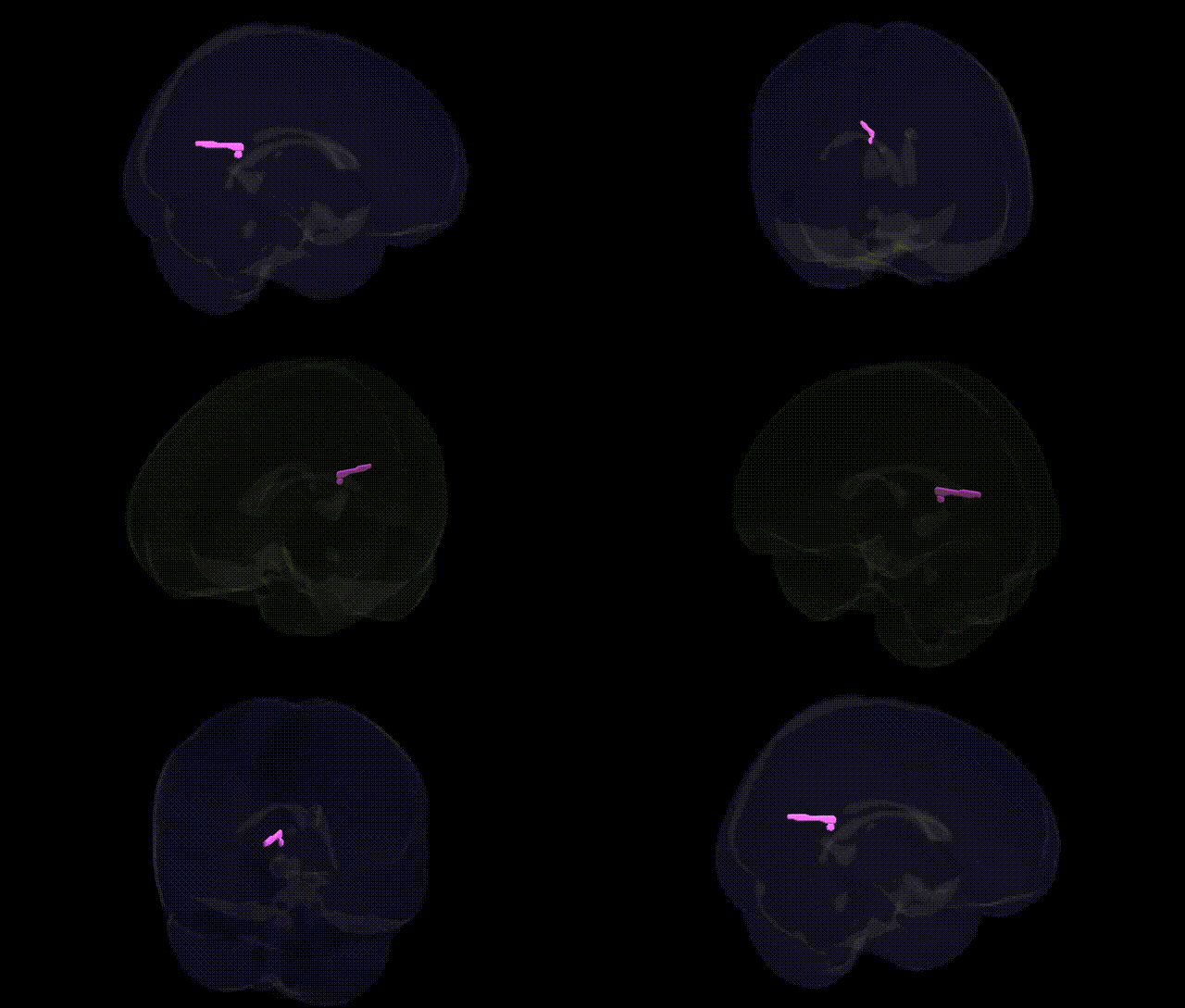
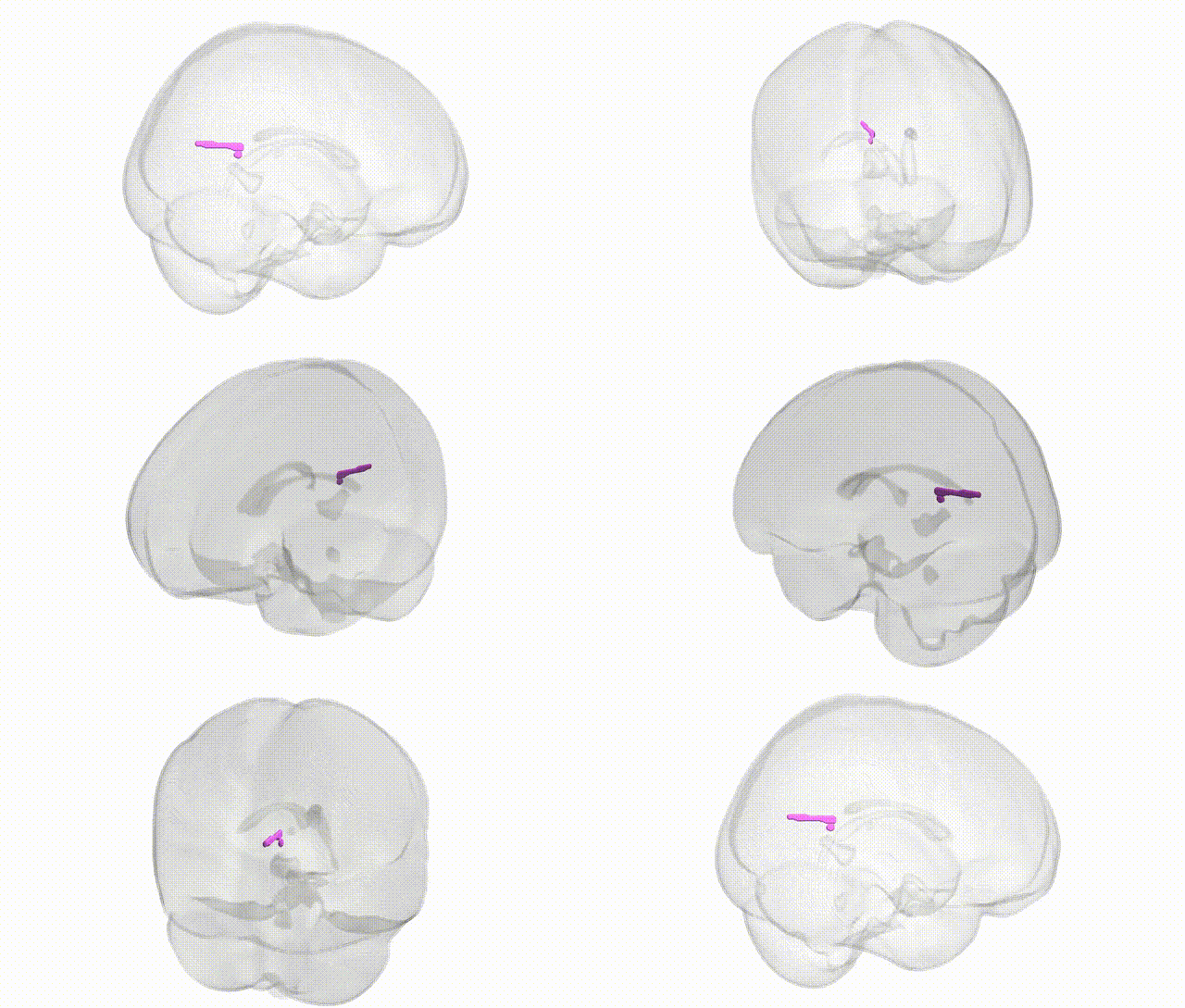
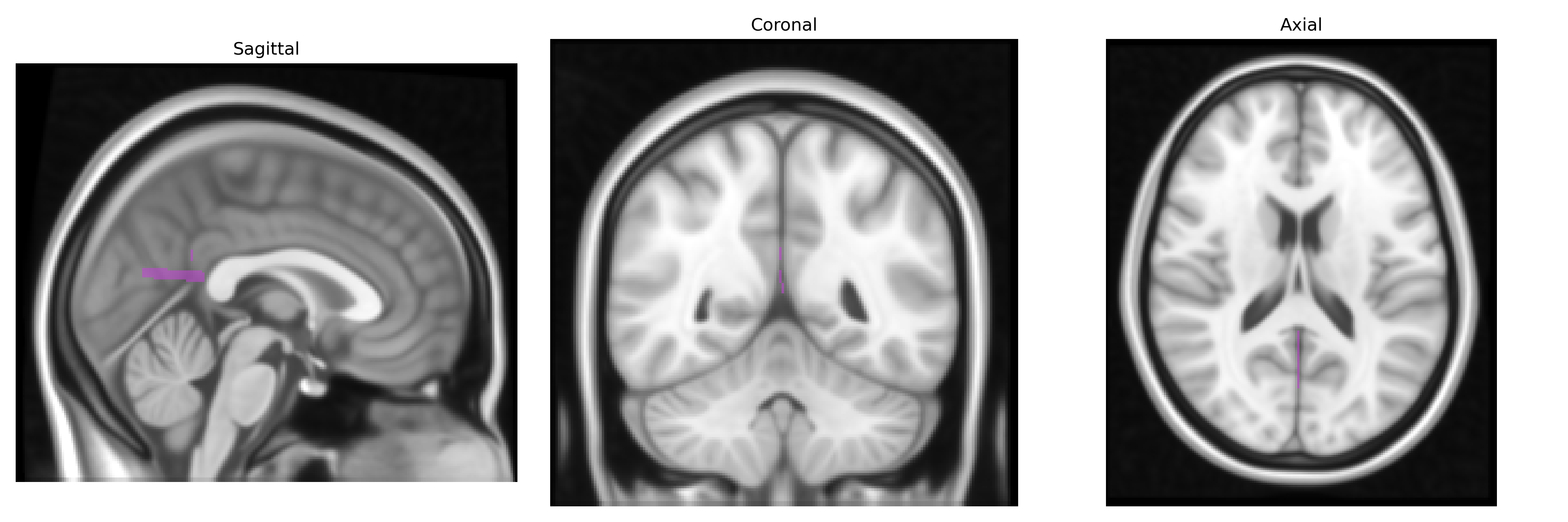
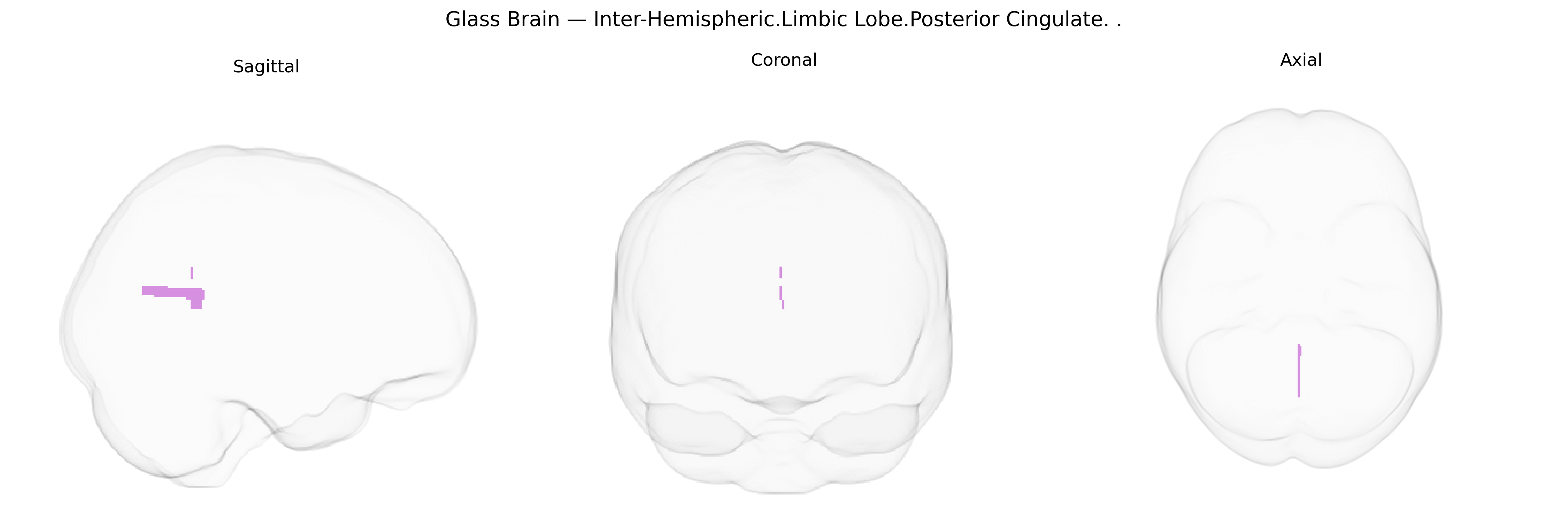
The bilateral Inter-Hemispheric Limbic Lobe Posterior Cingulate corresponds primarily to the posterior portion of the cingulate gyrus, situated along the medial surface of the cerebral hemispheres within the limbic lobe and bordering the precuneus and retrosplenial cortex. This region is strongly implicated in the default mode network, contributing to internally directed cognition, autobiographical memory retrieval, and self-referential processing. It is also involved in visuospatial orientation, emotional evaluation of stimuli, and integration of multimodal information relevant to conscious awareness. Metabolically active at rest and showing deactivation during many goal-directed tasks, the posterior cingulate is a key hub in large-scale brain networks and is notably affected in neurodegenerative conditions such as Alzheimer’s disease. Posterior cingulate cortex
The bilateral Inter-Hemispheric Limbic Lobe Posterior Cingulate, corresponding to the posterior cingulate cortex (PCC) in the Talairach 1 mm atlas, has been repeatedly implicated in genetic and GWAS-based imaging studies that link variation in this region’s structure and function to neuropsychiatric and neurodegenerative disorders. Twin and family studies indicate substantial heritability of PCC gray-matter volume, cortical thickness, and functional connectivity, particularly within the default mode network, with SNP-based heritability estimates often in the moderate range. Large-scale imaging–genetics consortia (e.g., ENIGMA, UK Biobank) have reported associations between common variants in genes related to synaptic plasticity, myelination, and neurodevelopment (such as pathways involving APOE, CLU, and other Alzheimer’s disease–related genes) and PCC volume or metabolism, consistent with the region’s sensitivity to early amyloid and tau pathology. GWAS of Alzheimer’s disease, schizophrenia, major depressive disorder, and autism spectrum traits have identified polygenic risk scores that correlate with altered PCC structure and default mode connectivity, with some studies linking PCC alterations to risk loci in genes involved in glutamatergic transmission, immune regulation, and calcium signaling. Additionally, genetic variants associated with cognitive performance, mind-wandering, and self-referential processing show effects on PCC activation patterns in task-based fMRI, highlighting the PCC as a convergence point where polygenic influences on cognition, affect, and neurodegenerative risk manifest in a specific limbic–default network hub.
Overview generated by GPT-4o (2026).
Region ID: 710
Hemisphere: bilateral
Atlas: Talairach labels 1mm
Full Quality Version: Download MP4
Full Quality Version: Download MP4
Wali Sidiqyar*, Gaurav Rudravaram*, Elyssa M. McMaster, Trent M. Schwartz, Adam M. Saunders, Kurt G. Schilling, Bennett A. Landman "Introducing SPINS: A Shared Public Visualization Library of Neuroanatomical Structures." Medical Imaging with Deep Learning- short paper
This resource is licensed under CC0 1.0 Universal (Public Domain).