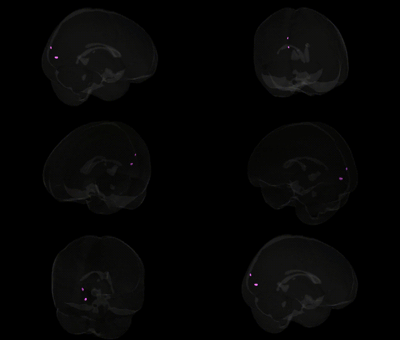
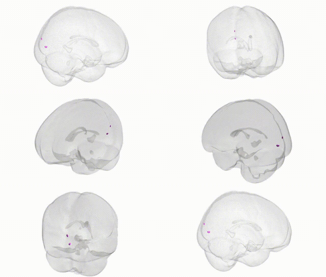
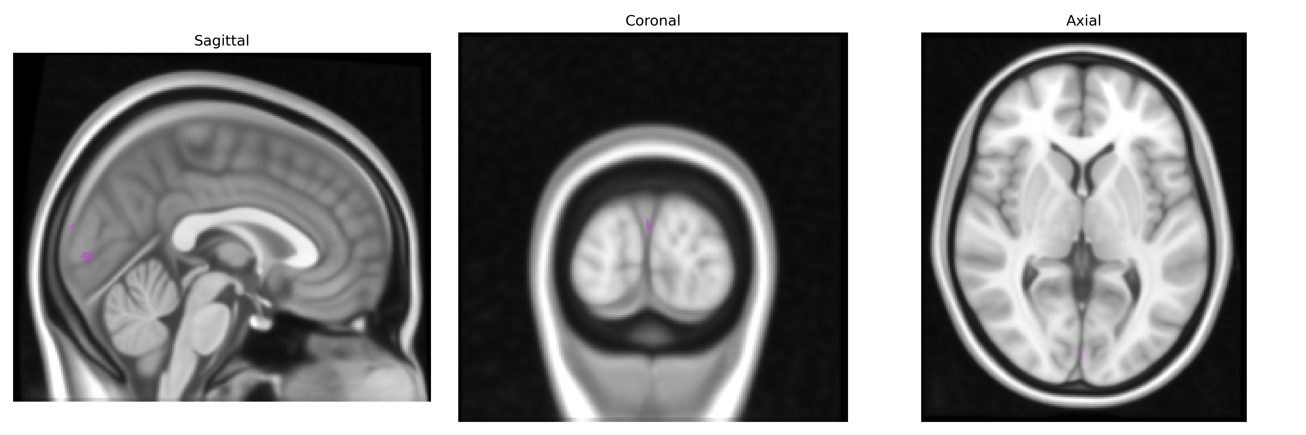
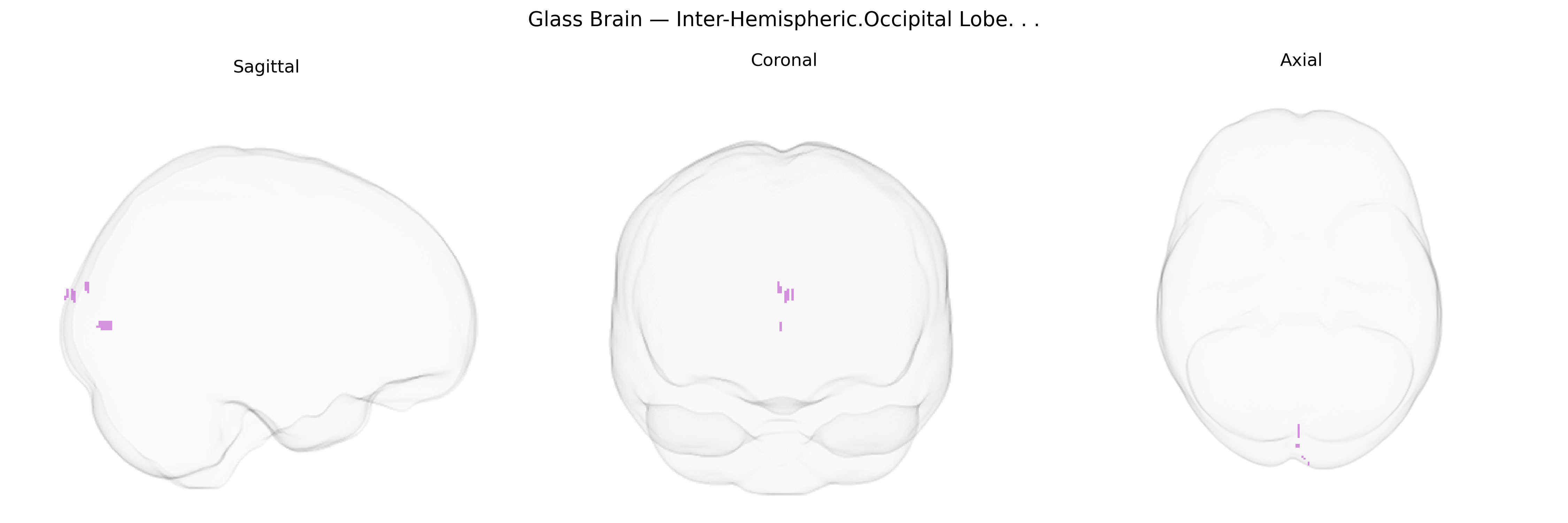
The bilateral inter-hemispheric occipital lobe refers to the medial portions of the occipital cortex that lie along and across the longitudinal fissure, encompassing regions from both the left and right hemispheres that participate in early and higher-order visual processing. These areas include midline visual cortices such as the cuneus and lingual gyrus and are involved in processing basic visual features (orientation, contrast, motion), integrating binocular input, and contributing to visual spatial organization and representation of the central visual field. The inter-hemispheric aspect emphasizes commissural connections via the splenium of the corpus callosum, which coordinate visual information between the two occipital lobes and support coherent perception across the visual field. No direct link exists for “bilateral inter-hemispheric occipital lobe”; a closely related structure is the Occipital lobe.
The bilateral inter-hemispheric occipital lobe, encompassing midline primary and secondary visual cortices, has been implicated in several genetic and GWAS findings that link visual processing and higher-order cognitive functions to specific loci and pathways. Variants in genes involved in synaptic development and axon guidance (such as those in the netrin, semaphorin, and Eph/ephrin families) have been repeatedly associated with structural measures of occipital cortex (including cortical thickness, surface area, and folding) in large neuroimaging-genetics consortia, while polygenic scores for general cognitive ability, educational attainment, and intracranial volume show correlations with occipital morphology and midline visual network connectivity. GWAS of brain functional networks and resting-state connectivity identify common variants influencing the strength and organization of visual networks, including interhemispheric occipital connections via the splenium of the corpus callosum, with enriched signals in genes related to myelination and oligodendrocyte biology (for example, within or near MBP and other myelin genes). In neuropsychiatric genetics, risk variants for schizophrenia, bipolar disorder, autism spectrum disorder, and major depression show distributed effects that include reduced occipital cortical thickness and altered visual network connectivity, often mediated through broadly expressed neurodevelopmental and synaptic genes (such as CACNA1C, GRIN2A, and multiple glutamatergic and GABAergic pathway genes) that influence visual cortex circuitry. Visual cortex–related genetic associations also emerge in disorders with strong sensory components, such as migraine with aura and photosensitive epilepsy, where GWAS implicate ion channel, neurotransmitter, and vascular-related genes that modulate occipital excitability and interhemispheric synchronization. Collectively, current evidence suggests that genetic influences on the inter-hemispheric occipital region are highly polygenic, shared across multiple cognitive and psychiatric traits, and concentrated in pathways regulating neurodevelopment, synaptic signaling, and myelination, rather than being driven by region-specific loci unique to this Talairach-defined area.
Overview generated by GPT-4o (2026).
Region ID: 547
Hemisphere: bilateral
Atlas: Talairach labels 1mm
Full Quality Version: Download MP4
Full Quality Version: Download MP4
Wali Sidiqyar*, Gaurav Rudravaram*, Elyssa M. McMaster, Trent M. Schwartz, Adam M. Saunders, Kurt G. Schilling, Bennett A. Landman "Introducing SPINS: A Shared Public Visualization Library of Neuroanatomical Structures." Medical Imaging with Deep Learning- short paper
This resource is licensed under CC0 1.0 Universal (Public Domain).