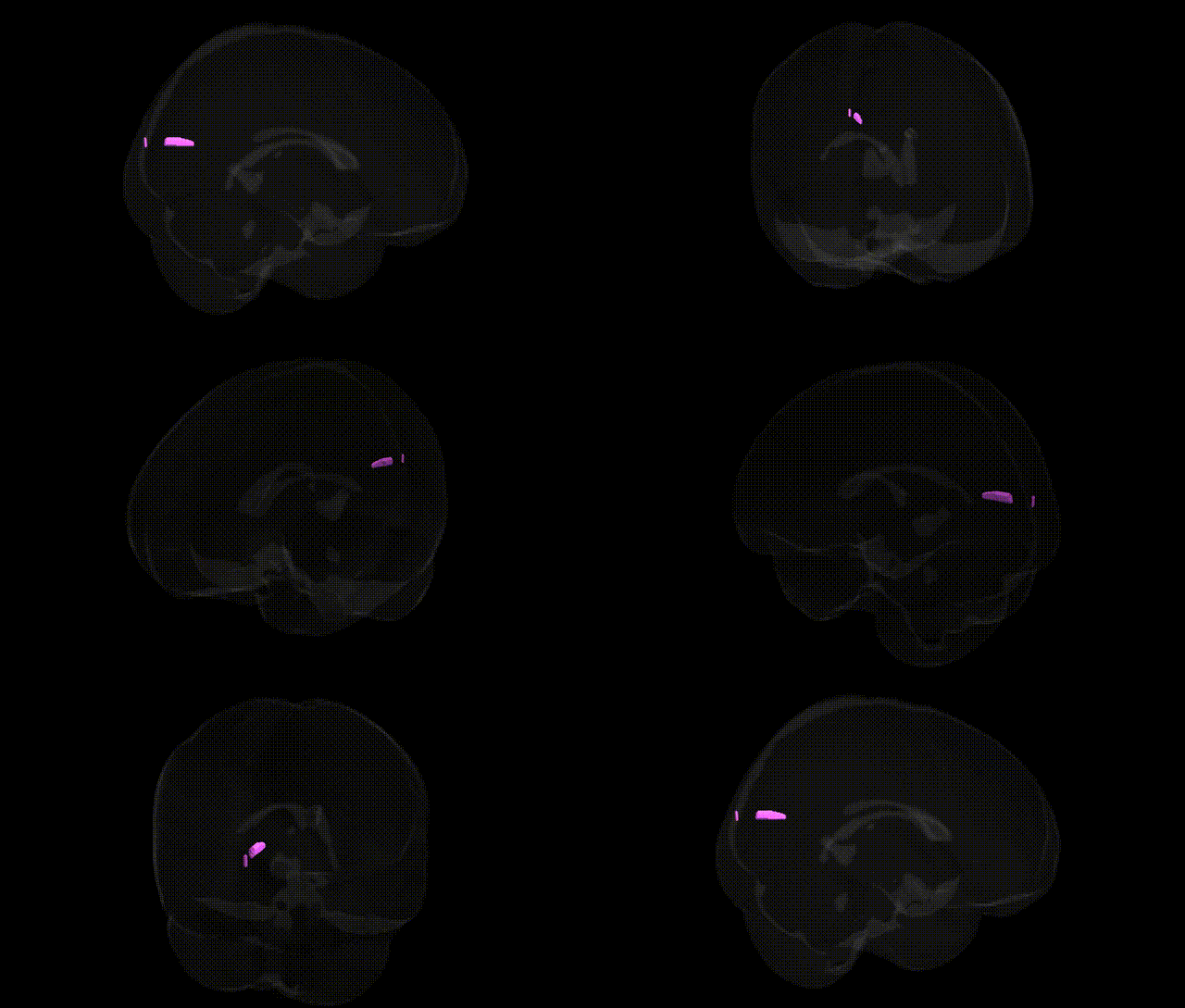
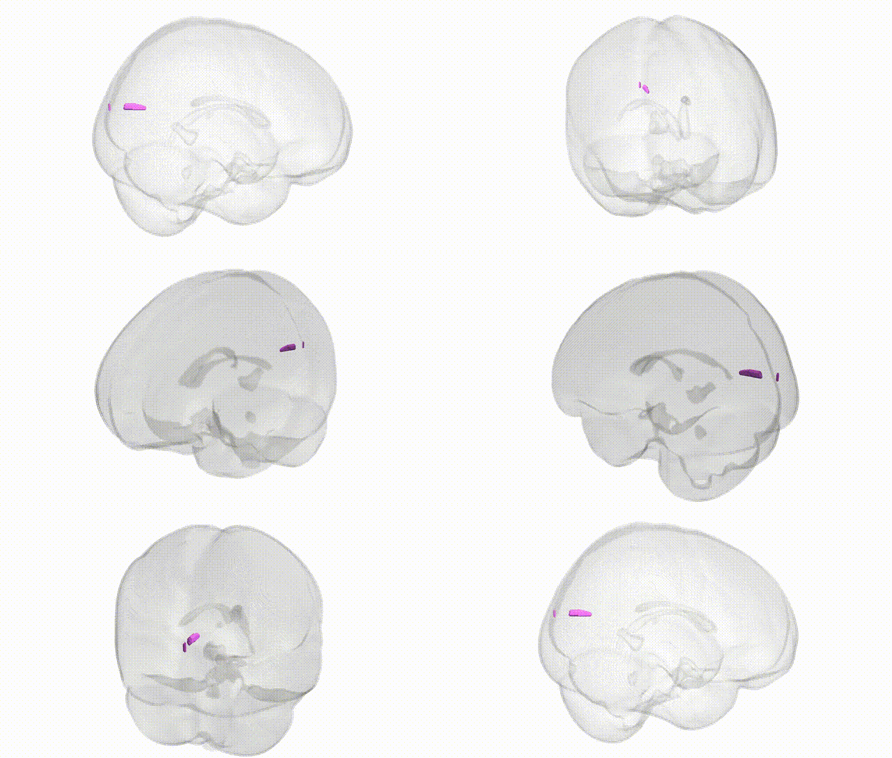
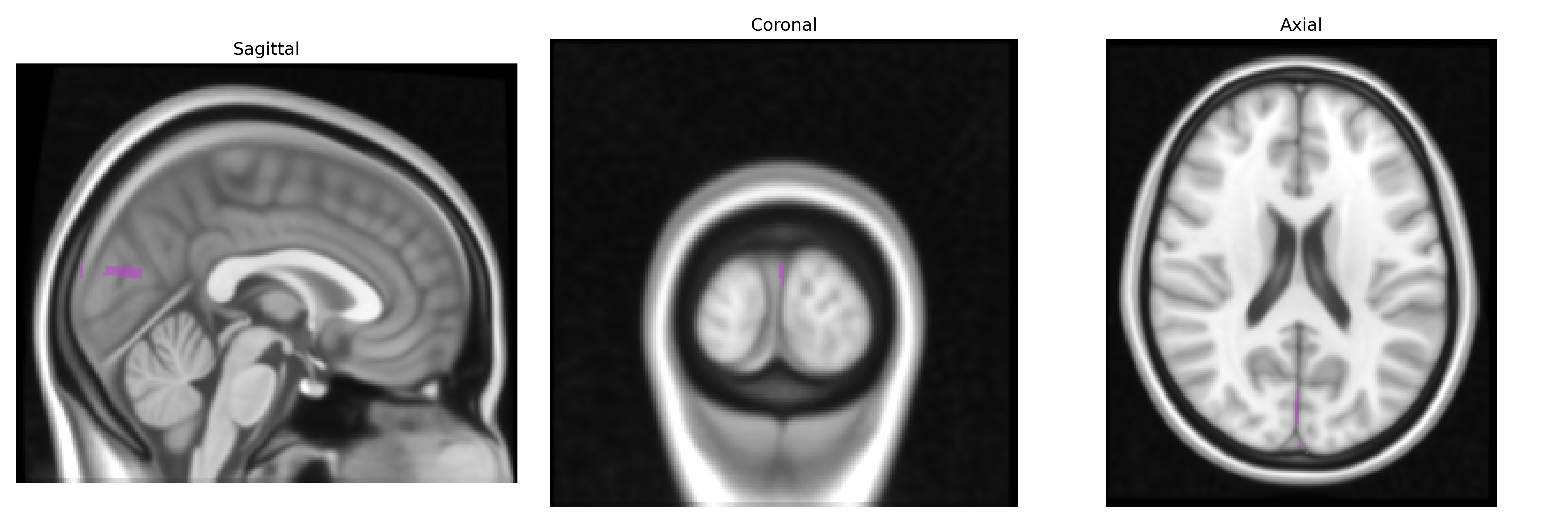
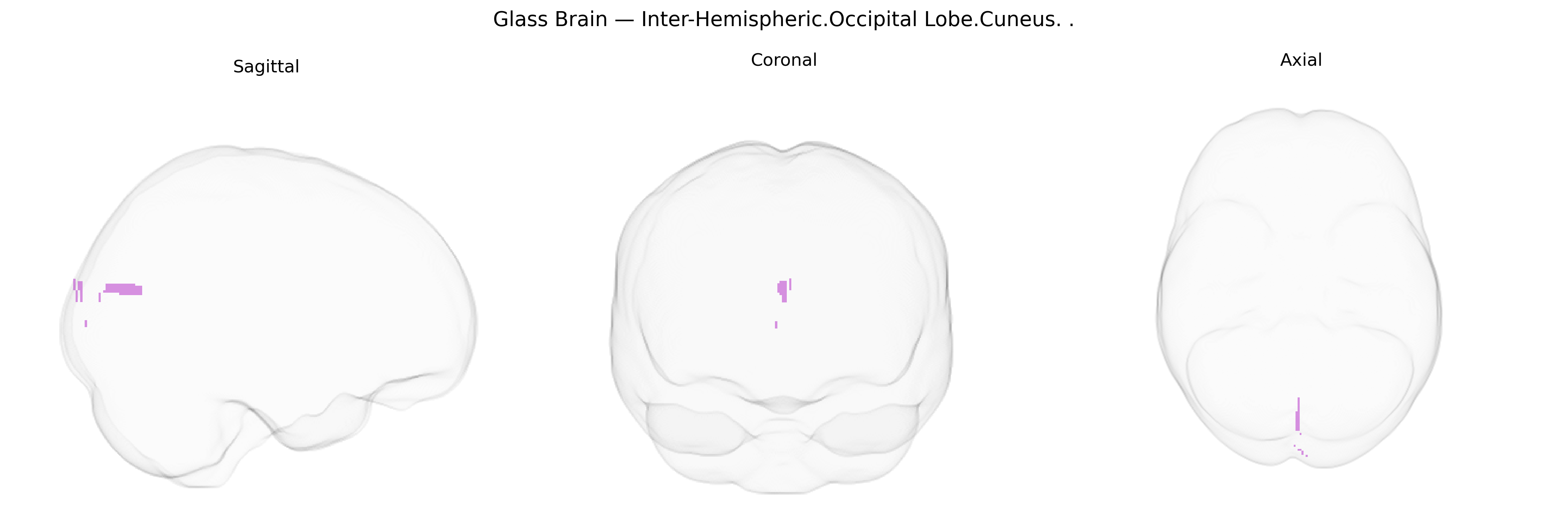
The bilateral inter-hemispheric occipital lobe cuneus refers to the medial portion of the occipital cortex in both hemispheres that lies along the longitudinal fissure, forming a wedge-shaped region (the cuneus) bounded superiorly by the parieto-occipital sulcus and inferiorly by the calcarine sulcus. This region is a key component of the primary and secondary visual cortices, receiving highly organized retinotopic input from the lateral geniculate nucleus of the thalamus and participating in early-stage processing of basic visual features such as orientation, spatial frequency, contrast, and motion. The cuneus contributes to the integration of visual information for spatial orientation, visual attention, and higher-order visual functions, and is interconnected with other occipital, parietal, and frontal visual association areas within dorsal and ventral visual processing streams. Cuneus
The bilateral interhemispheric occipital lobe cuneus (Talairach 1 mm atlas) is a primary visual and higher-order visual association region whose structure and function show substantial heritability in imaging–genetics and GWAS studies, with common variants in genes related to neurodevelopment, synaptic function, and myelination repeatedly implicated. Large-scale brain MRI GWAS (e.g., ENIGMA, UK Biobank) have linked cuneus cortical thickness, surface area, and volume to loci involving cell-adhesion and axon-guidance genes (such as CADM2, CNTNAP2, and ROBO family genes), as well as neurodevelopmental regulators (including genes in Wnt and Notch pathways); many of these loci overlap with those associated with general cognitive ability, educational attainment, and intracranial volume. Functional and resting-state GWAS and polygenic score analyses show that variation in cuneus activity and connectivity is enriched for schizophrenia, major depression, autism spectrum disorder, and ADHD polygenic risk, consistent with frequent cuneus involvement in case–control neuroimaging studies of these disorders. Visual cortex–focused GWAS and candidate gene analyses also report associations between cuneus morphometry or activation and genetic risk for migraine, especially variants near TRPM8 and other pain- and sensory-related genes, as well as genes linked to ocular traits and refractive error. Collectively, genetic findings suggest that the cuneus is a heritable hub where variants influencing early visual processing, attention, and large-scale brain networks contribute to vulnerability for neuropsychiatric and neurodevelopmental disorders, as well as to normal variability in visual and cognitive performance.
Overview generated by GPT-4o (2026).
Region ID: 543
Hemisphere: bilateral
Atlas: Talairach labels 1mm
Full Quality Version: Download MP4
Full Quality Version: Download MP4
Wali Sidiqyar*, Gaurav Rudravaram*, Elyssa M. McMaster, Trent M. Schwartz, Adam M. Saunders, Kurt G. Schilling, Bennett A. Landman "Introducing SPINS: A Shared Public Visualization Library of Neuroanatomical Structures." Medical Imaging with Deep Learning- short paper
This resource is licensed under CC0 1.0 Universal (Public Domain).