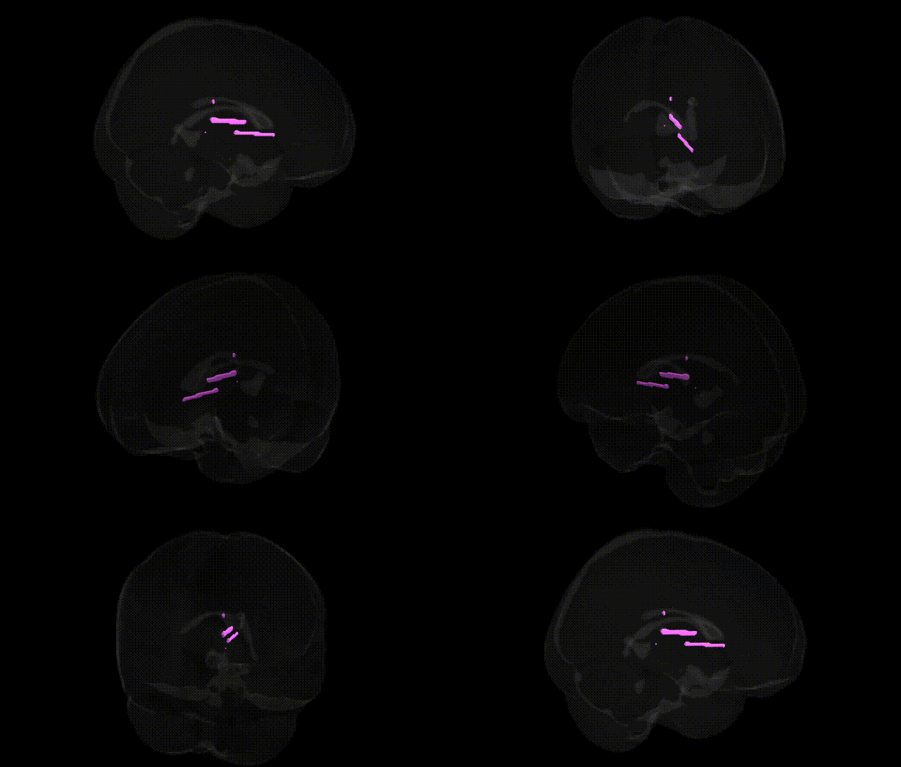
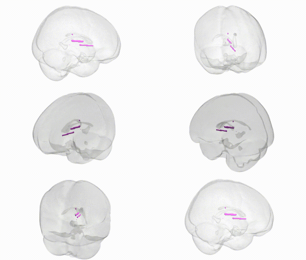
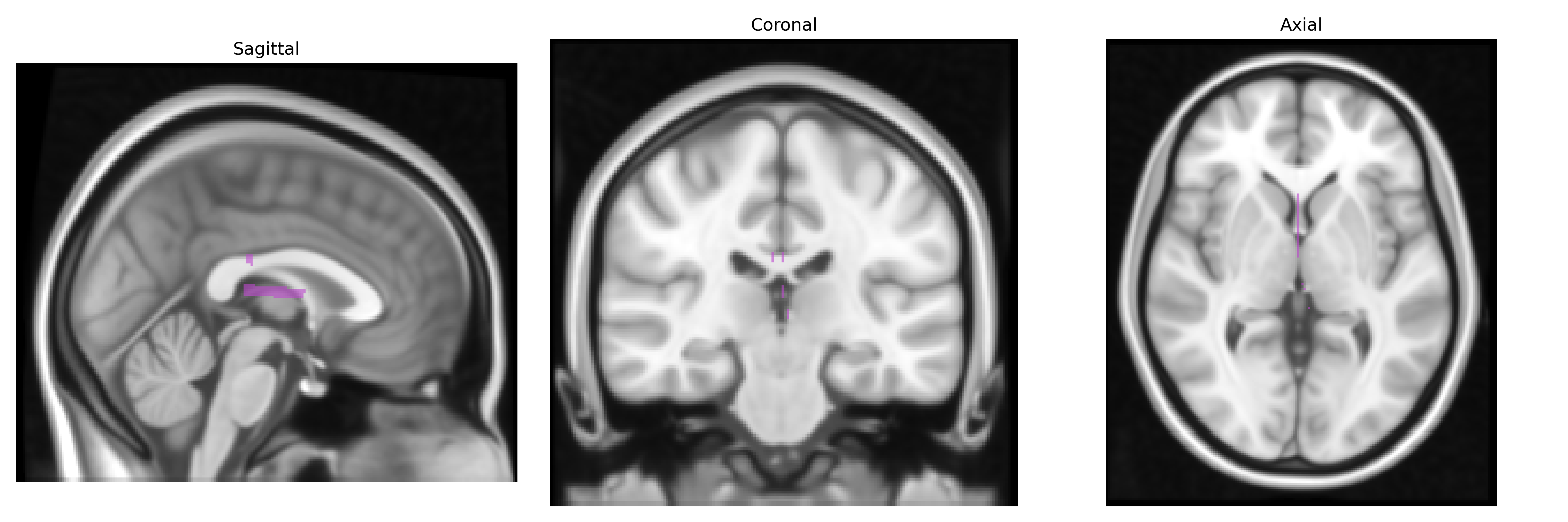
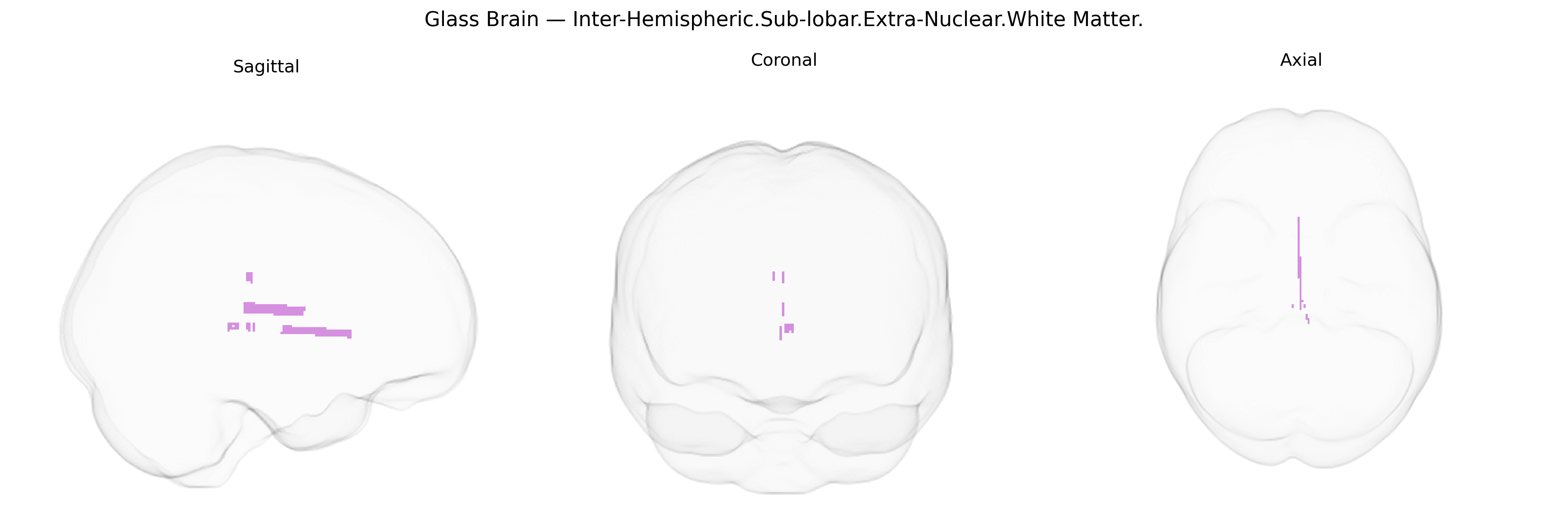
The bilateral Inter-Hemispheric Sub-lobar Extra-Nuclear White Matter, as defined in the Talairach 1 mm atlas, refers to deep, midline white matter tracts situated between the cerebral hemispheres, inferior to the cortical lobes and outside the major nuclear (gray matter) collections such as the basal ganglia and thalamus. This region comprises interhemispheric fiber systems—including portions of commissural pathways—that facilitate communication between homologous and associated cortical and subcortical areas across the two hemispheres, contributing to integration of sensory, motor, and higher-order cognitive functions. Structurally, it is embedded within the broader subcortical white matter and lies in close relation to major midline structures such as the corpus callosum and cingulum bundle. There is no direct link for this specific Talairach-defined label; a related structure is the White matter.
Genetic associations specific to the Talairach “bilateral Inter-Hemispheric.Sub-lobar.Extra-Nuclear.White Matter” label have not been directly reported, but convergent evidence from GWAS of commissural and interhemispheric white matter—particularly involving the corpus callosum and adjacent midline tracts—implicates multiple gene pathways and neuropsychiatric traits. Large-scale imaging-genetics studies (e.g., ENIGMA, UK Biobank) show that common variants in genes involved in axon guidance (e.g., ROBO1/ROBO2, DCC, SLIT2), myelination and oligodendrocyte function (e.g., MBP, MAG, MOG, PLP1, ERBB4), and neurodevelopmental patterning (e.g., L1CAM, NCAM1) are associated with diffusion MRI measures (fractional anisotropy, mean diffusivity) in midline white-matter tracts that correspond anatomically to this region. These genetic influences overlap with loci linked to neurodevelopmental and psychiatric disorders, including schizophrenia, bipolar disorder, autism spectrum disorder, ADHD, and major depression, where altered callosal or interhemispheric white-matter integrity is frequently reported; some of these loci (such as variants near CACNA1C, ZNF804A, and MIR137 regions) show parallel effects on both disorder risk and white-matter microstructure. Additional associations involve genes implicated in neurodegeneration and small-vessel disease (e.g., APOE for Alzheimer’s disease, HTRA1 and COL4A1-related pathways for leukoaraiosis and white-matter hyperintensities), where GWAS of white-matter hyperintensity burden and diffuse white-matter changes highlight vascular, inflammatory, and extracellular-matrix genes that influence midline and periventricular white matter. Overall, the genetic architecture of this interhemispheric sublobar white-matter territory reflects a complex interplay of neurodevelopmental, myelination, and vascular pathways that also contribute to risk for psychiatric, cognitive, and cerebrovascular phenotypes, even though no study has isolated this Talairach-defined region as a unique genetic target.
Overview generated by GPT-4o (2026).
Region ID: 621
Hemisphere: bilateral
Atlas: Talairach labels 1mm
Full Quality Version: Download MP4
Full Quality Version: Download MP4
Wali Sidiqyar*, Gaurav Rudravaram*, Elyssa M. McMaster, Trent M. Schwartz, Adam M. Saunders, Kurt G. Schilling, Bennett A. Landman "Introducing SPINS: A Shared Public Visualization Library of Neuroanatomical Structures." Medical Imaging with Deep Learning- short paper
This resource is licensed under CC0 1.0 Universal (Public Domain).