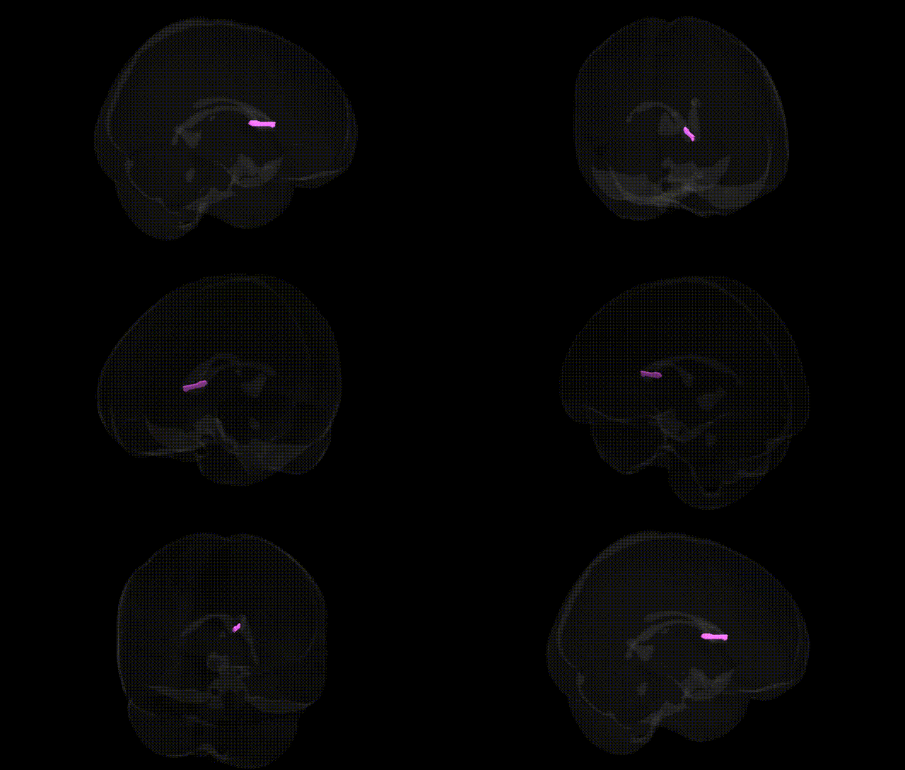
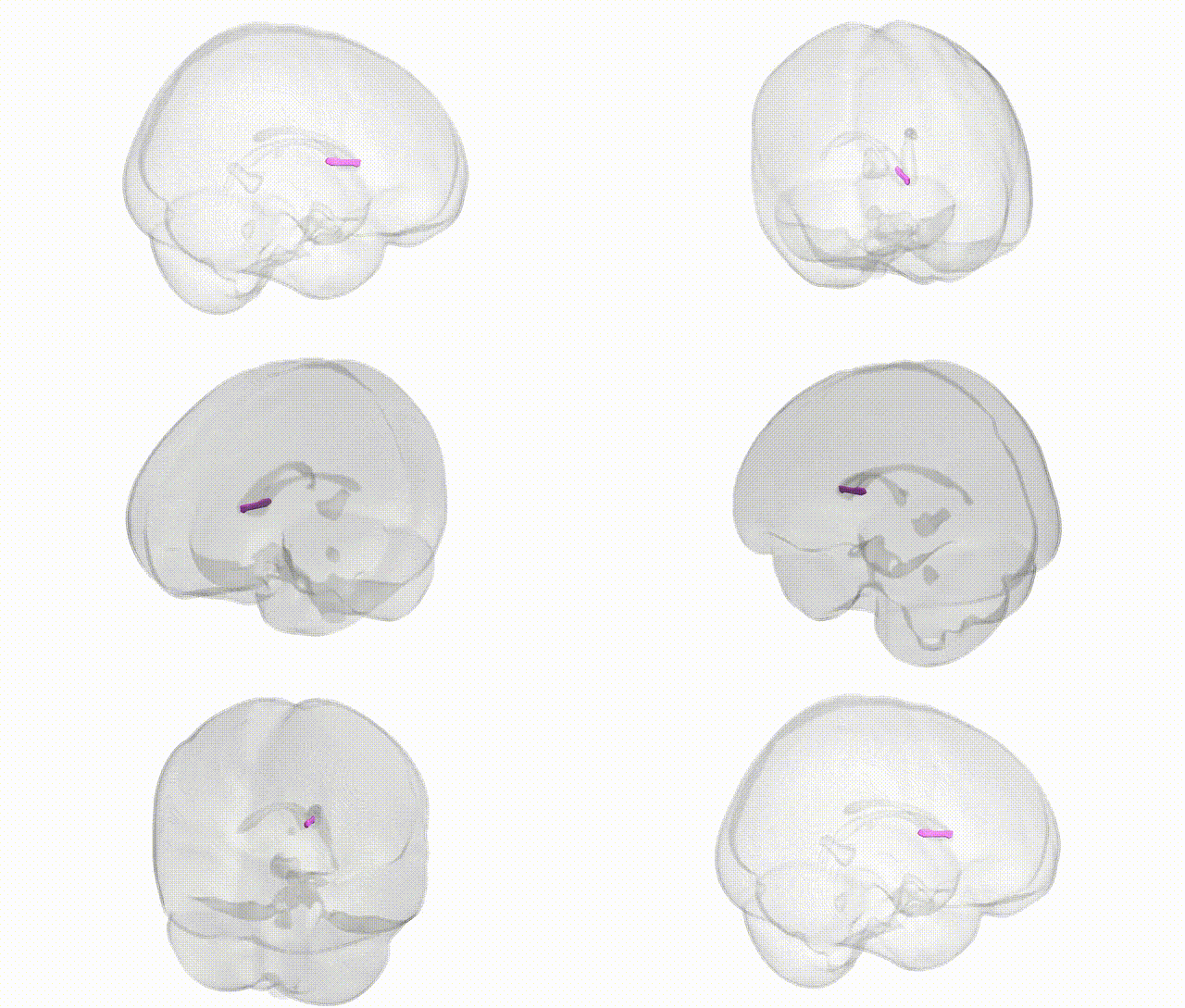
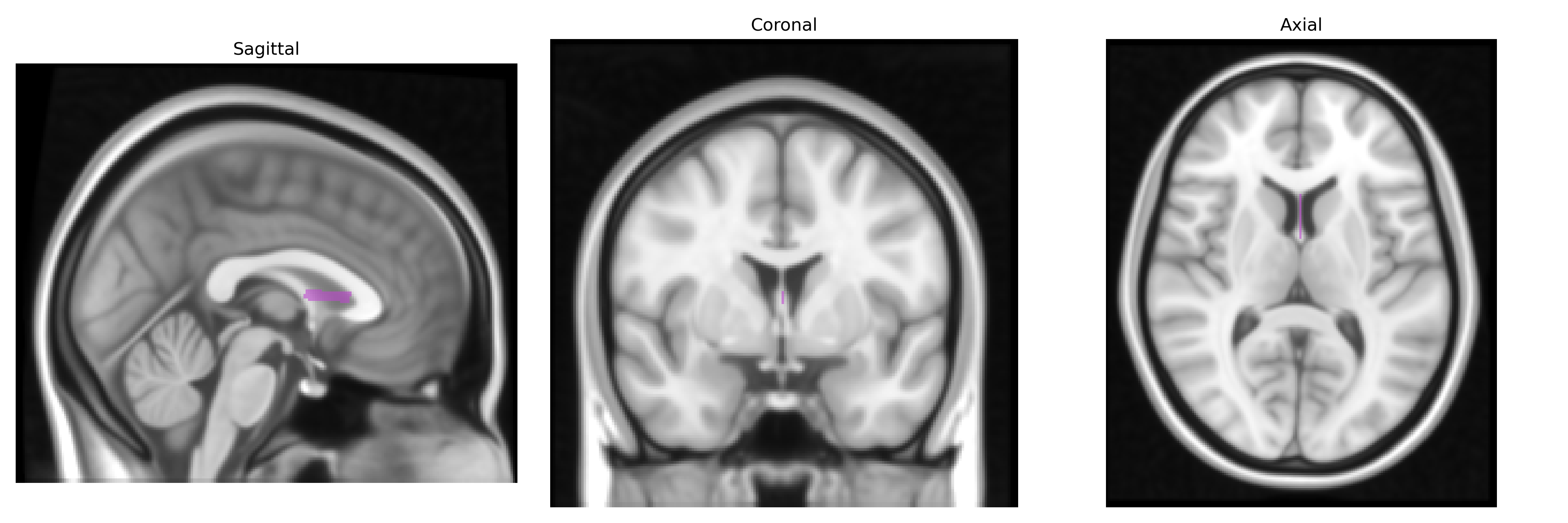
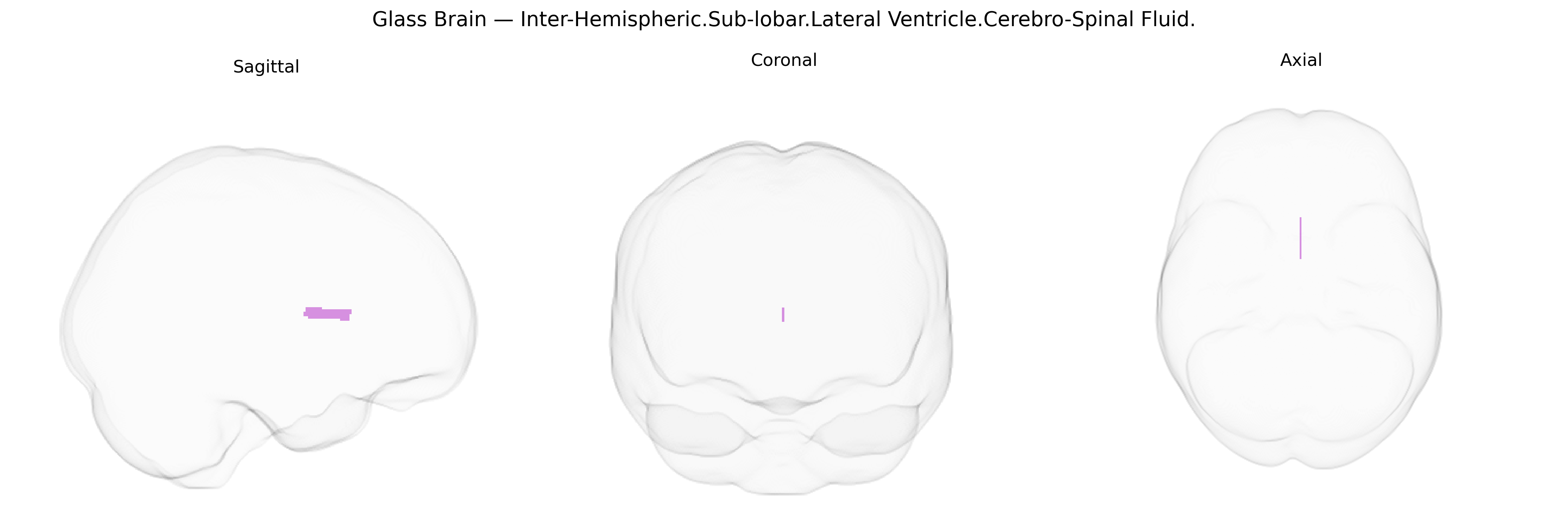
The bilateral Inter-Hemispheric Sub-lobar Lateral Ventricle Cerebro-Spinal Fluid region corresponds to the cerebrospinal fluid (CSF) occupying the lateral ventricles, a paired C-shaped cavity system located deep within each cerebral hemisphere. The lateral ventricles consist of anterior (frontal), body (parietal), posterior (occipital), and inferior (temporal) horns, and are lined by ependymal cells, forming part of the ventricular system that circulates CSF produced mainly by the choroid plexus. This CSF-filled space cushions the brain, helps maintain intracranial pressure, removes metabolic waste, and participates in homeostatic regulation of the central nervous system. There is no direct Wikipedia article for this exact Talairach label; a closely related structure is the Lateral ventricle.
The lateral ventricles, which contain cerebrospinal fluid and serve as a proxy for global and regional brain atrophy, have been extensively examined in imaging-genetics and GWAS studies, with numerous loci implicated in inter-individual variation of ventricular volume, including variants in and near genes such as APOE (particularly ε4, via its strong association with Alzheimer’s disease and neurodegeneration), MAPT, CLU, PICALM, and BIN1, as well as genes involved in neurodevelopment, axonal guidance, and extracellular matrix organization (e.g., variants near WNT signaling genes and collagen-related loci). Large-scale consortia (e.g., ENIGMA, UK Biobank, CHARGE) have repeatedly identified robust associations between lateral ventricular enlargement and polygenic risk for Alzheimer’s disease, schizophrenia, bipolar disorder, major depressive disorder, and other neuropsychiatric conditions, as well as with general cognitive decline and aging-related traits. GWAS of ventricular volume highlight high heritability and implicate pathways related to neuroinflammation, synaptic structure, and myelination, suggesting that genetic factors influencing neurodegenerative and neurodevelopmental processes contribute to the observed inter-hemispheric and sublobar CSF-space differences captured by this Talairach-defined Inter-Hemispheric.Sub-lobar.Lateral Ventricle.Cerebro-Spinal Fluid region.
Overview generated by GPT-4o (2026).
Region ID: 744
Hemisphere: bilateral
Atlas: Talairach labels 1mm
Full Quality Version: Download MP4
Full Quality Version: Download MP4
Wali Sidiqyar*, Gaurav Rudravaram*, Elyssa M. McMaster, Trent M. Schwartz, Adam M. Saunders, Kurt G. Schilling, Bennett A. Landman "Introducing SPINS: A Shared Public Visualization Library of Neuroanatomical Structures." Medical Imaging with Deep Learning- short paper
This resource is licensed under CC0 1.0 Universal (Public Domain).