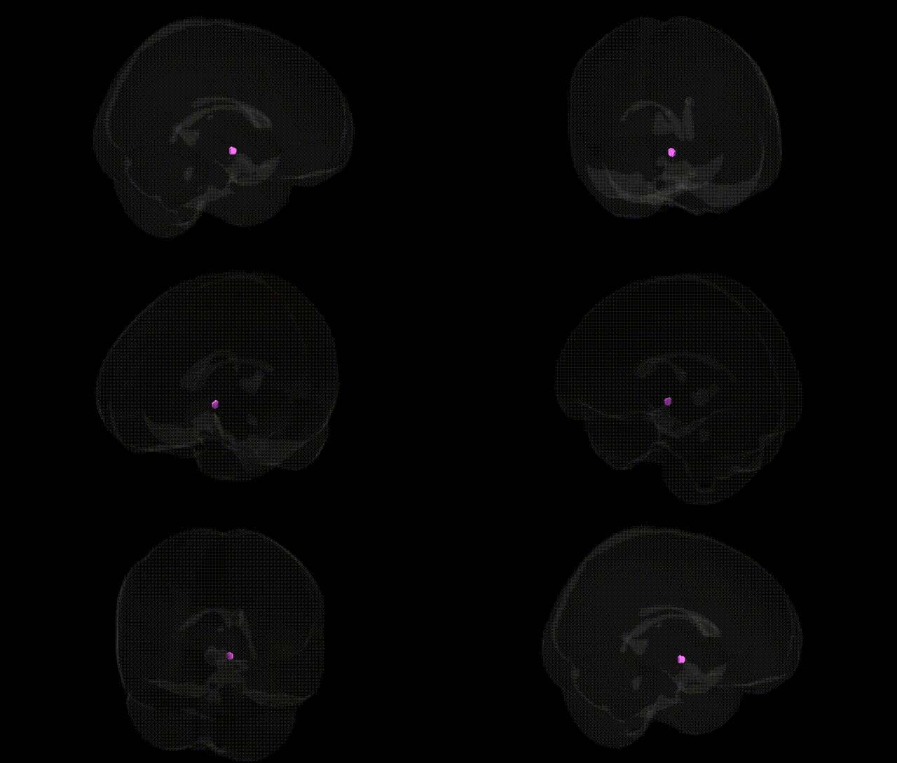
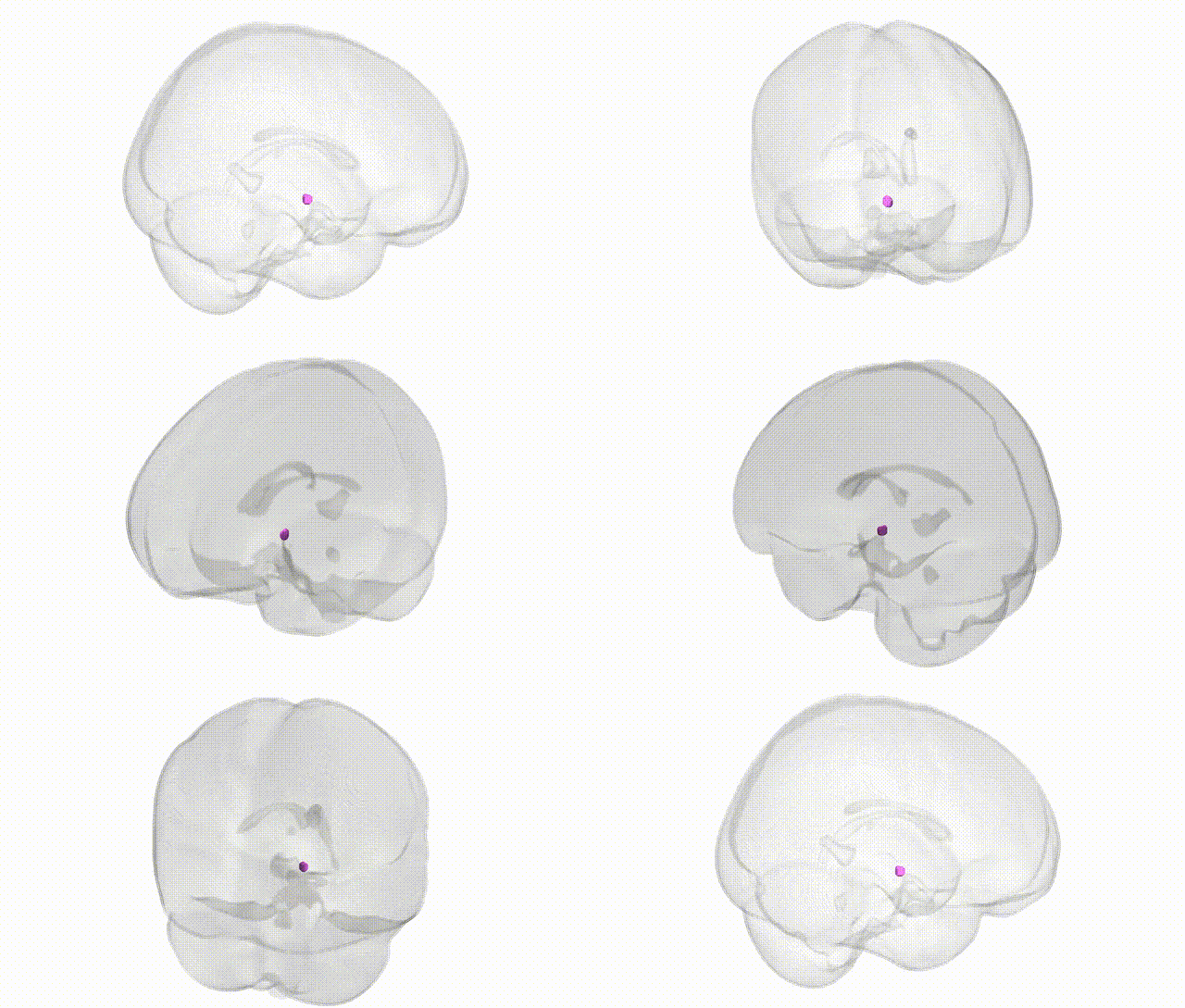
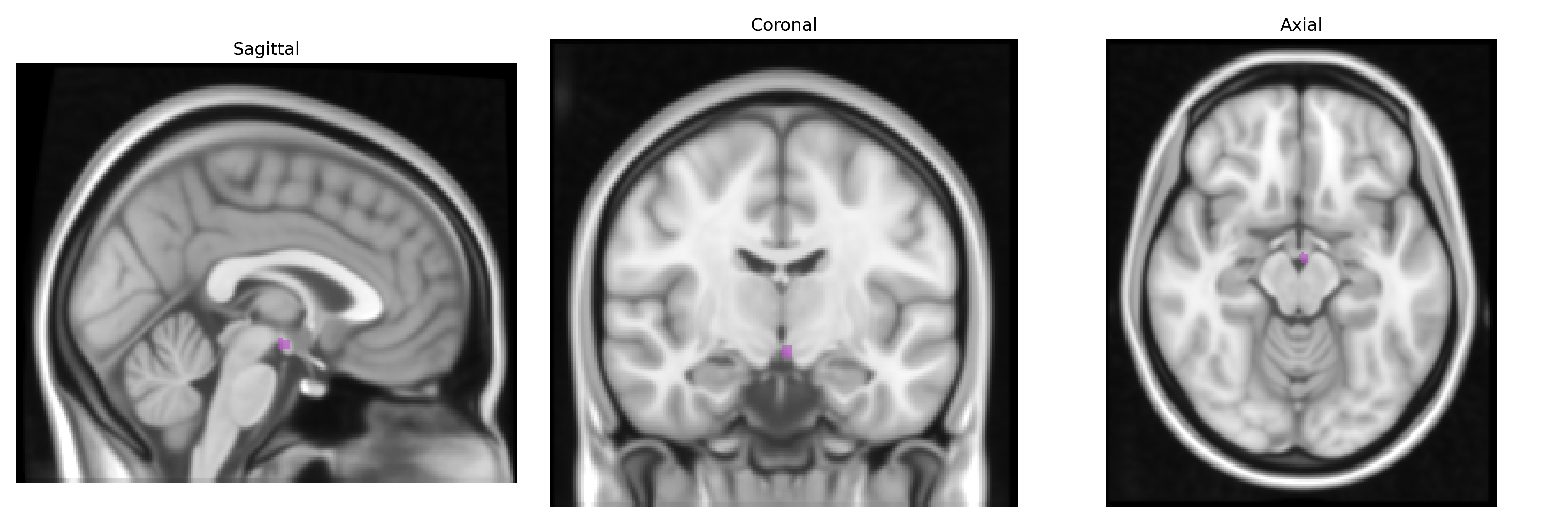
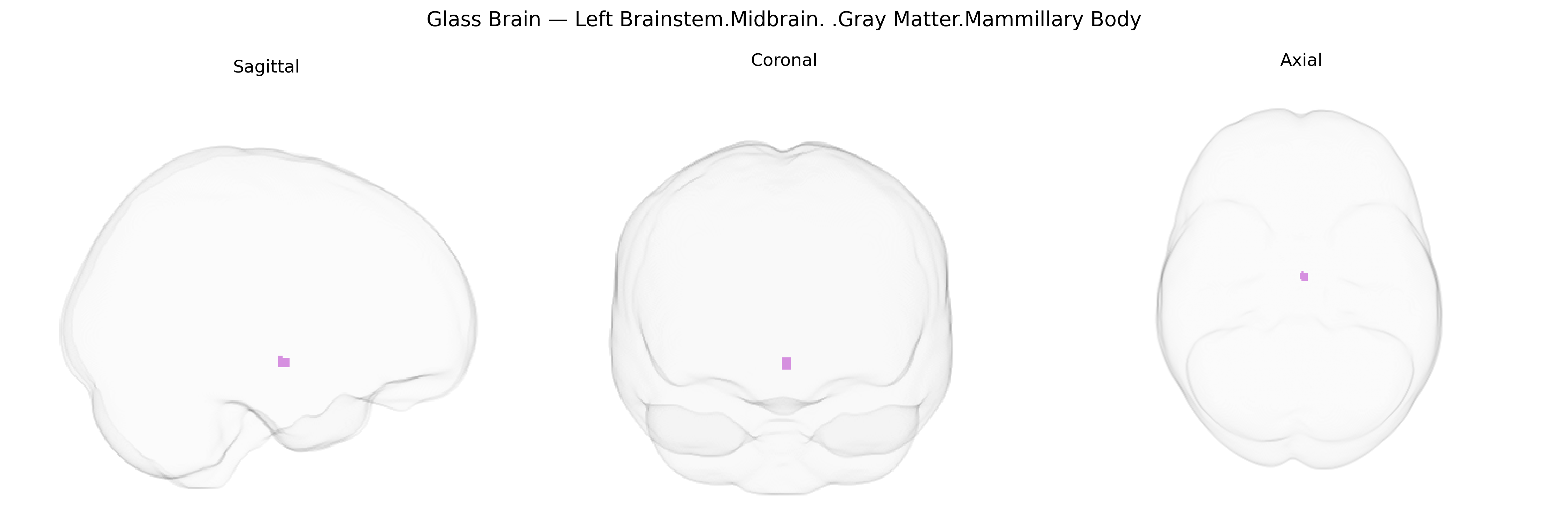
The bilateral Left Brainstem.Midbrain.Gray Matter.Mammillary Body corresponds to the mammillary bodies, paired hypothalamic structures located on the ventral surface of the diencephalon near the midbrain junction. These rounded gray matter nuclei are integral components of the limbic system and form a key relay in the Papez circuit, receiving major input from the hippocampal formation via the fornix and projecting primarily to the anterior thalamic nuclei through the mammillothalamic tract. They play a critical role in episodic memory processing, particularly in recollective and spatial aspects of memory, and are notably vulnerable in thiamine deficiency, where lesions contribute to the amnestic syndromes characteristic of Wernicke–Korsakoff syndrome. As part of the broader hypothalamic network, they also participate in integrating mnemonic and autonomic information. Mammillary body
The mammillary bodies, small hypothalamic structures within the midbrain/brainstem region, have been most prominently implicated in genetic studies of memory circuits and neuropsychiatric disease rather than in large, region-specific GWAS directly targeting this Talairach-defined area; instead, evidence comes from disorders in which mammillary body atrophy, volume change, or functional disruption is part of a broader circuit pathology. Genetic variants in genes involved in thiamine transport and metabolism (such as SLC19A2 and other thiamine-related pathways) are indirectly linked through susceptibility to Wernicke–Korsakoff syndrome and related amnestic conditions that prominently affect the mammillary bodies, while broader polygenic risk for schizophrenia, bipolar disorder, and major depression has been associated with structural and connectivity alterations in Papez circuit components that include this region. Imaging–genetics and large-scale ENIGMA-type studies of subcortical and limbic structures report that common variants near genes influencing neurodevelopment, synaptic function, and myelination (for example, loci near MAPT, NTRK2, BDNF, and other neurotrophic or axonal guidance genes) are associated with limbic and diencephalic volumes, with the mammillary bodies typically analyzed as part of hypothalamic or hippocampal–diencephalic networks rather than as isolated ROIs. Rare familial and syndromic conditions affecting midline diencephalic development (such as some cases of septo-optic dysplasia or midline malformation syndromes involving genes like HESX1 or SOX2) can include mammillary body abnormalities, further underscoring a genetic contribution to its structure, but explicit GWAS hits naming “mammillary body” as a primary locus remain scarce, and current genetic associations are largely inferred from its participation in memory, diencephalic, and limbic circuitry affected by polygenic neuropsychiatric and neurodegenerative risk.
Overview generated by GPT-4o (2026).
Region ID: 354
Hemisphere: bilateral
Atlas: Talairach labels 1mm
Full Quality Version: Download MP4
Full Quality Version: Download MP4
Wali Sidiqyar*, Gaurav Rudravaram*, Elyssa M. McMaster, Trent M. Schwartz, Adam M. Saunders, Kurt G. Schilling, Bennett A. Landman "Introducing SPINS: A Shared Public Visualization Library of Neuroanatomical Structures." Medical Imaging with Deep Learning- short paper
This resource is licensed under CC0 1.0 Universal (Public Domain).