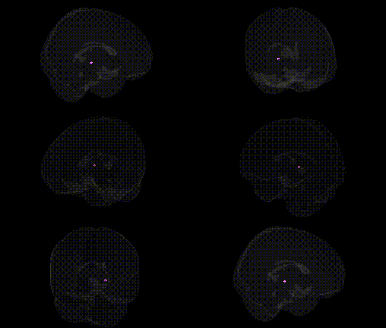
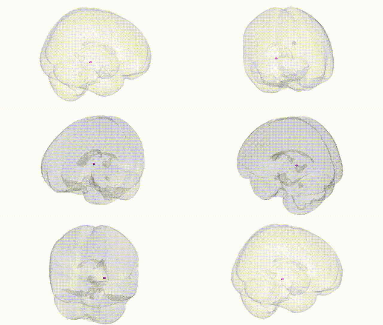
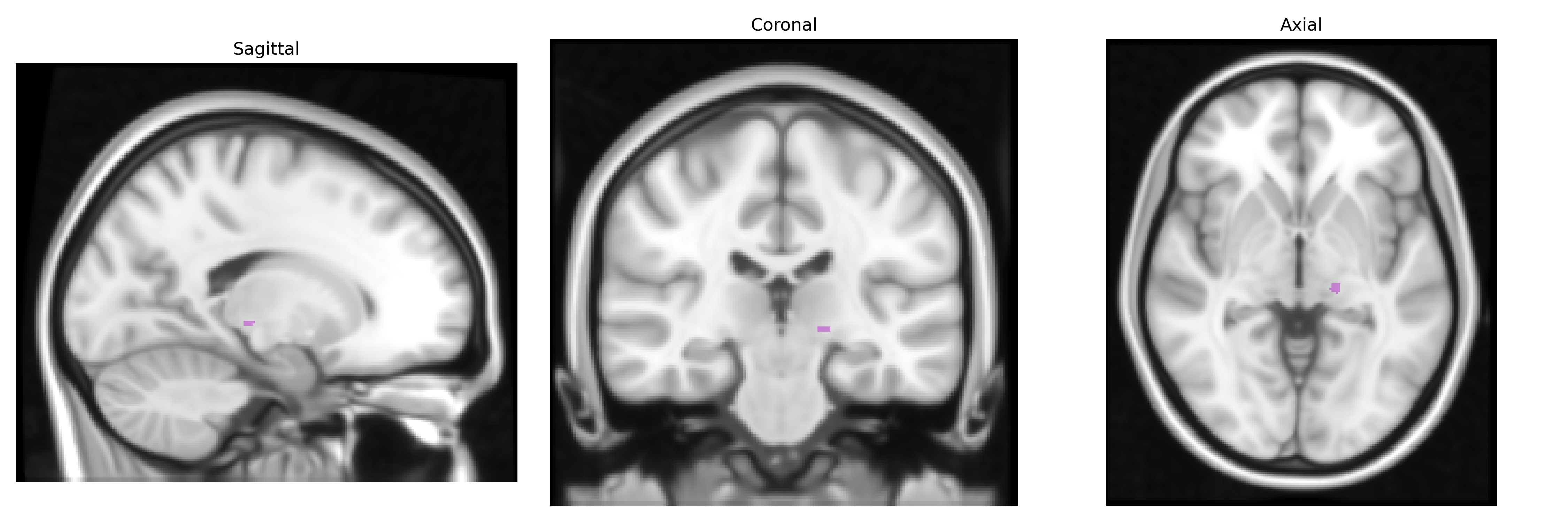
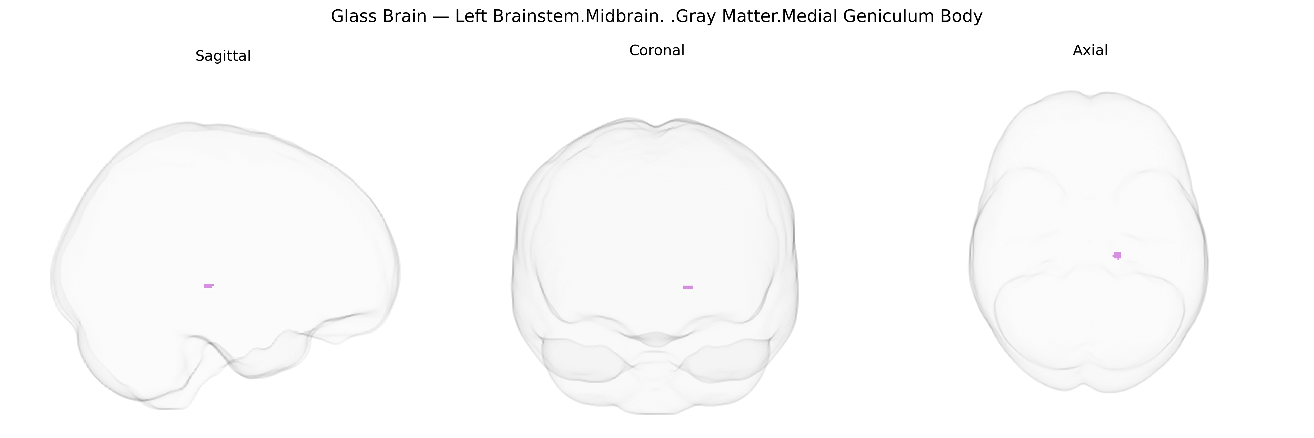
The bilateral Left Brainstem Midbrain Gray Matter Medial Geniculum Body corresponds to the medial geniculate body (MGB), a key thalamic relay nucleus for the auditory system situated near the midbrain–thalamus junction. It receives ascending auditory input primarily from the inferior colliculus and projects to the primary auditory cortex (Heschl’s gyrus), participating in the processing of sound frequency, intensity, and temporal patterns, as well as in aspects of auditory attention. Histologically, the MGB is subdivided into ventral, dorsal, and medial divisions, each with distinct cytoarchitecture and connectivity, supporting specialized functions such as precise tonotopic mapping (ventral division) and more integrative, multimodal processing (dorsal and medial divisions). In Talairach-based neuroimaging atlases, activation within this region is often interpreted in the context of auditory perception, sound localization, and higher-order auditory cognition. Medial geniculate nucleus
The medial geniculate body, a key auditory thalamic relay closely integrated with midbrain brainstem pathways, has been implicated in several genetically influenced traits and disorders through MRI- and diffusion-based GWAS and imaging–genetics studies, although findings are relatively sparse compared with cortical regions. Large consortia such as ENIGMA and UK Biobank have identified heritable variation in thalamic nuclei volumes and microstructure, including the auditory/medial geniculate region, with associated loci enriched in neurodevelopmental and synaptic genes (for example, variants near genes involved in axon guidance, synaptic vesicle cycling, and neuronal differentiation), though most signals are not region-specific and reflect broader subcortical development. Genetic studies of tinnitus and hearing-related traits (e.g., GWAS in UK Biobank and HUNT) have highlighted loci in genes such as GJB2, PCDH20, and others that affect central auditory processing, with functional imaging suggesting altered medial geniculate activity in tinnitus, but direct, robust SNP associations to medial geniculate morphology or function remain limited. In neuropsychiatric disorders like schizophrenia and bipolar disorder, risk variants (e.g., in CACNA1C, GRIN2A, and other synaptic/ion-channel genes) have been associated with thalamocortical circuit abnormalities, and some structural and functional MRI studies report involvement of the medial geniculate body within disrupted auditory and salience networks, yet these links are typically inferred at the circuit level rather than via region-specific GWAS. Similarly, autism spectrum disorder and dyslexia have been connected to atypical auditory thalamic processing, with genetic risk concentrated in synaptic and transcriptional regulators (such as CNTNAP2, FOXP2-related networks, and chromatin-remodeling genes), but again without strong evidence for medial geniculate–specific genetic effects. Overall, the bilateral medial geniculate/midbrain gray matter region appears as a node in heritable auditory and thalamocortical circuits affected by polygenic variation, with current genetic associations pointing to general neurodevelopmental and synaptic pathways rather than clearly defined, region-specific risk alleles.
Overview generated by GPT-4o (2026).
Region ID: 498
Hemisphere: bilateral
Atlas: Talairach labels 1mm
Full Quality Version: Download MP4
Full Quality Version: Download MP4
Wali Sidiqyar*, Gaurav Rudravaram*, Elyssa M. McMaster, Trent M. Schwartz, Adam M. Saunders, Kurt G. Schilling, Bennett A. Landman "Introducing SPINS: A Shared Public Visualization Library of Neuroanatomical Structures." Medical Imaging with Deep Learning- short paper
This resource is licensed under CC0 1.0 Universal (Public Domain).