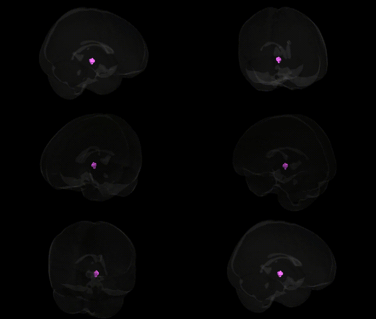
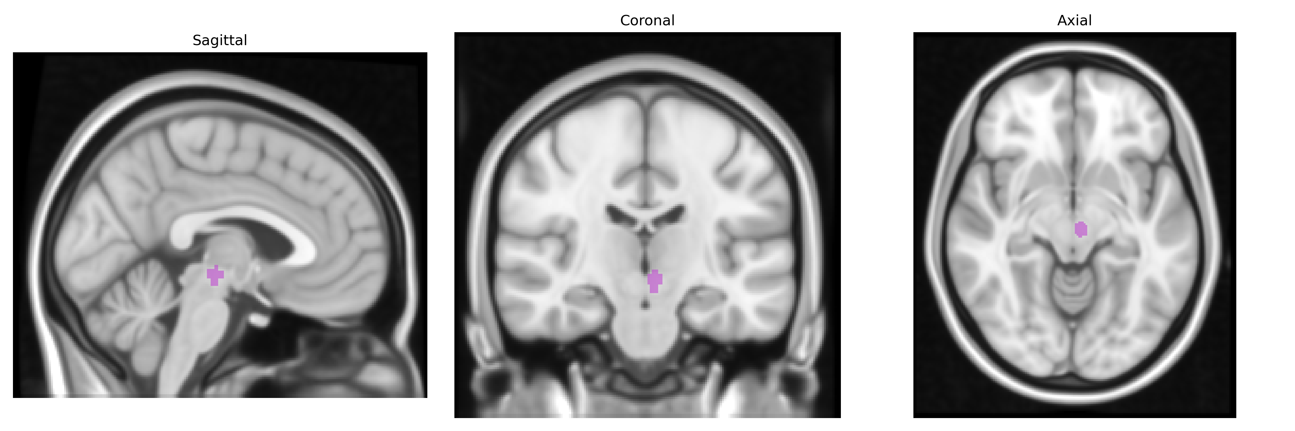
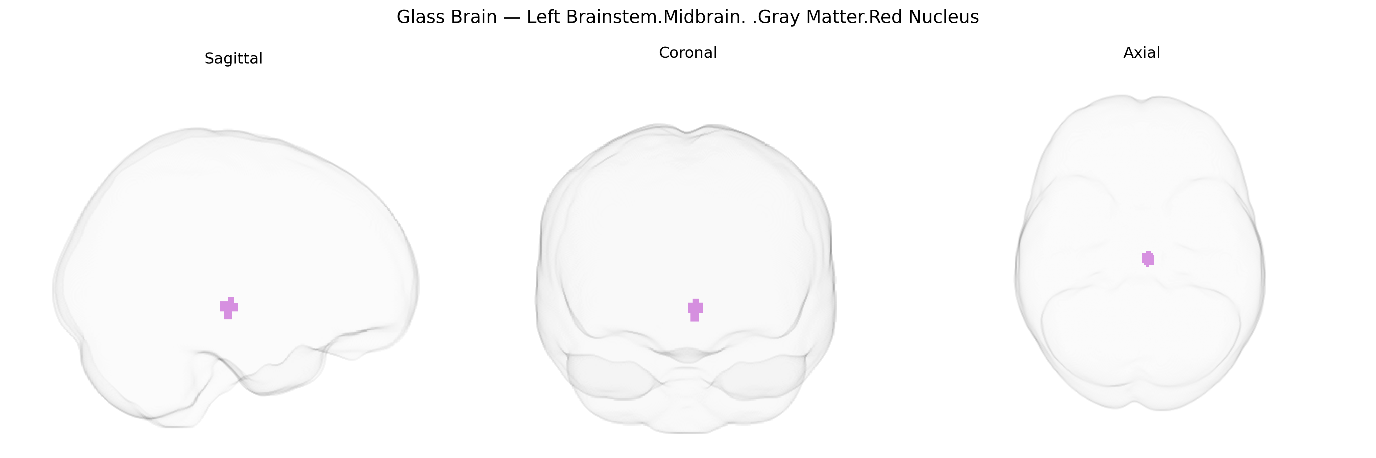
The bilateral Left Brainstem.Midbrain.Gray Matter.Red Nucleus corresponds to the paired red nuclei located in the rostral midbrain tegmentum, dorsal to the substantia nigra and medial to the cerebral peduncles, and is a key relay in motor coordination and motor learning. Each red nucleus receives major afferent input from the contralateral cerebellar nuclei (via the superior cerebellar peduncle) and the ipsilateral motor cortex, and sends efferent projections through the rubrospinal tract to the spinal cord and via rubro-olivary connections that form part of cerebello–olivo–rubral loops. Structurally, the red nucleus has a magnocellular part (more prominent in many non-human mammals) and a parvocellular part (well developed in humans) that is heavily involved in integrating cerebellar and cortical signals, particularly for distal limb movement control and fine motor adjustment; it also plays a role in motor adaptation and in shaping voluntary movement timing. The region is richly vascularized and pigmented (owing to iron-containing pigments), which gives it a reddish color in gross anatomical preparations and contributes to its conspicuous appearance in midbrain cross-sections. Red nucleus
The red nucleus, a midbrain gray matter structure implicated in motor coordination, has been indirectly linked to several genetic influences through imaging genetics and GWAS of brain morphology and related motor and psychiatric phenotypes, although few studies target it specifically in the Talairach 1 mm atlas framework. Variants in genes involved in neurodevelopment and axonal guidance (such as ROBO1/ROBO2, L1CAM, and other corticospinal and cerebellar pathway genes) have been associated with midbrain and brainstem volumetric differences, which encompass the red nucleus region, while genes influencing iron metabolism (e.g., HFE and other iron-regulatory loci) have been implicated in individual differences in iron-sensitive MRI contrast in midbrain nuclei that include the red nucleus. Polygenic risk scores for Parkinson’s disease and essential tremor, which involve red nucleus circuitry as part of cerebello-thalamo-cortical and rubro-spinal pathways, show associations with altered brainstem and midbrain structure and function, and GWAS of essential tremor and dystonia highlight loci related to synaptic transmission and cerebellar/midbrain development that likely affect this region’s integrity. Additionally, common variants associated with schizophrenia, major depression, and ADHD have been linked via large-scale imaging–genetic studies to altered midbrain volumes and functional connectivity in motor and reward circuits that include the red nucleus, suggesting that its structure and function may be modulated by widespread polygenic influences rather than a small number of region-specific variants.
Overview generated by GPT-4o (2026).
Region ID: 343
Hemisphere: bilateral
Atlas: Talairach labels 1mm
Full Quality Version: Download MP4
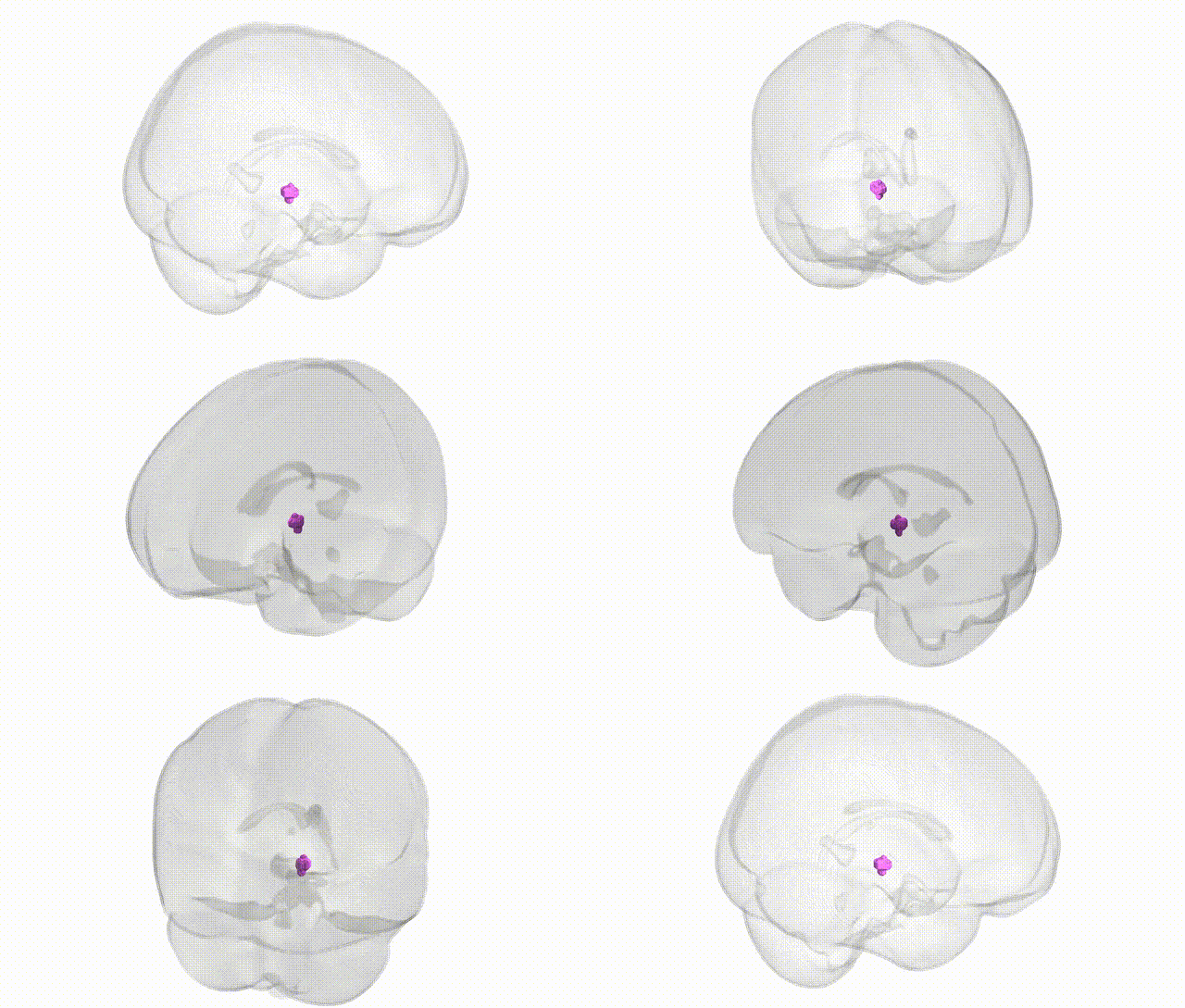
Full Quality Version: Download MP4
Wali Sidiqyar*, Gaurav Rudravaram*, Elyssa M. McMaster, Trent M. Schwartz, Adam M. Saunders, Kurt G. Schilling, Bennett A. Landman "Introducing SPINS: A Shared Public Visualization Library of Neuroanatomical Structures." Medical Imaging with Deep Learning- short paper
This resource is licensed under CC0 1.0 Universal (Public Domain).