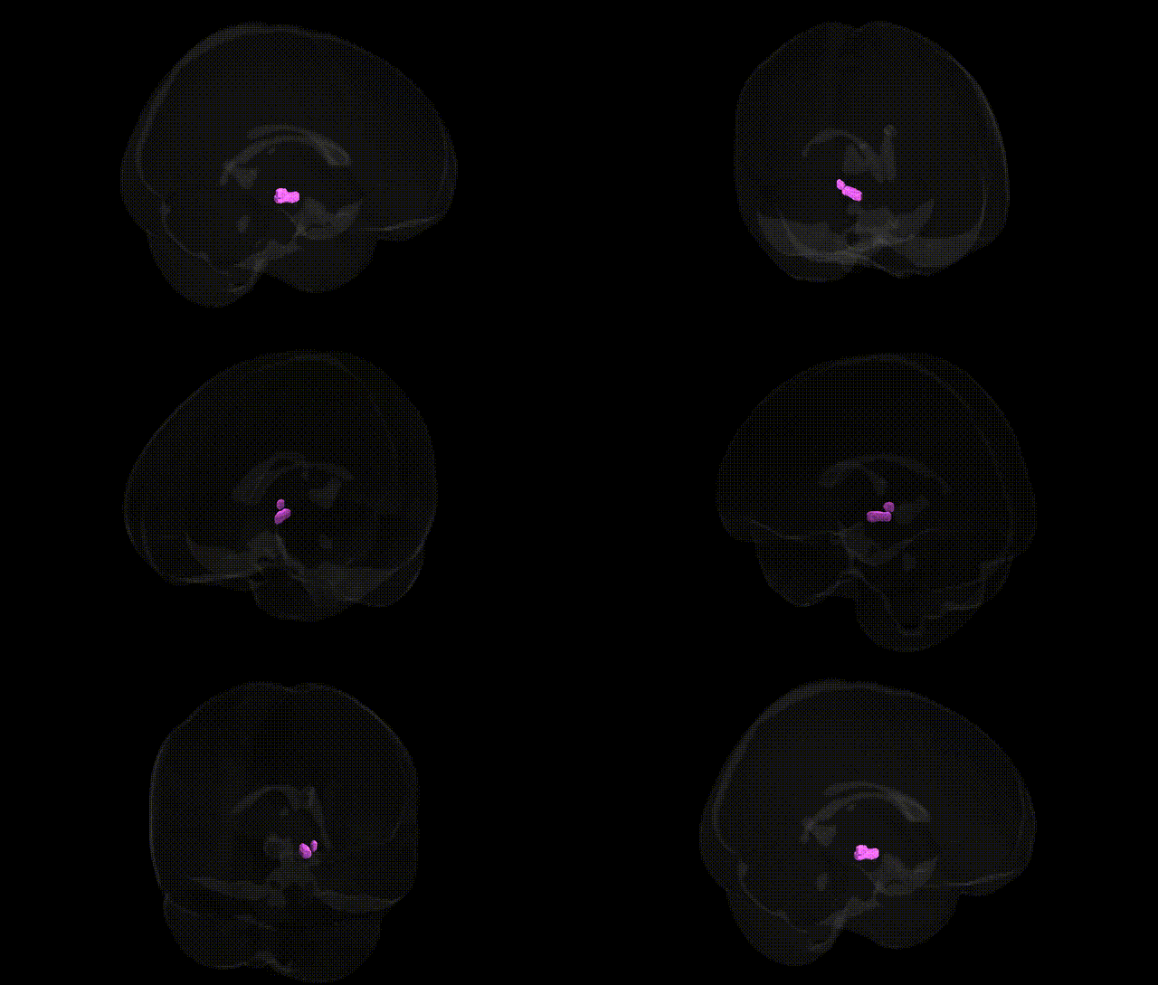
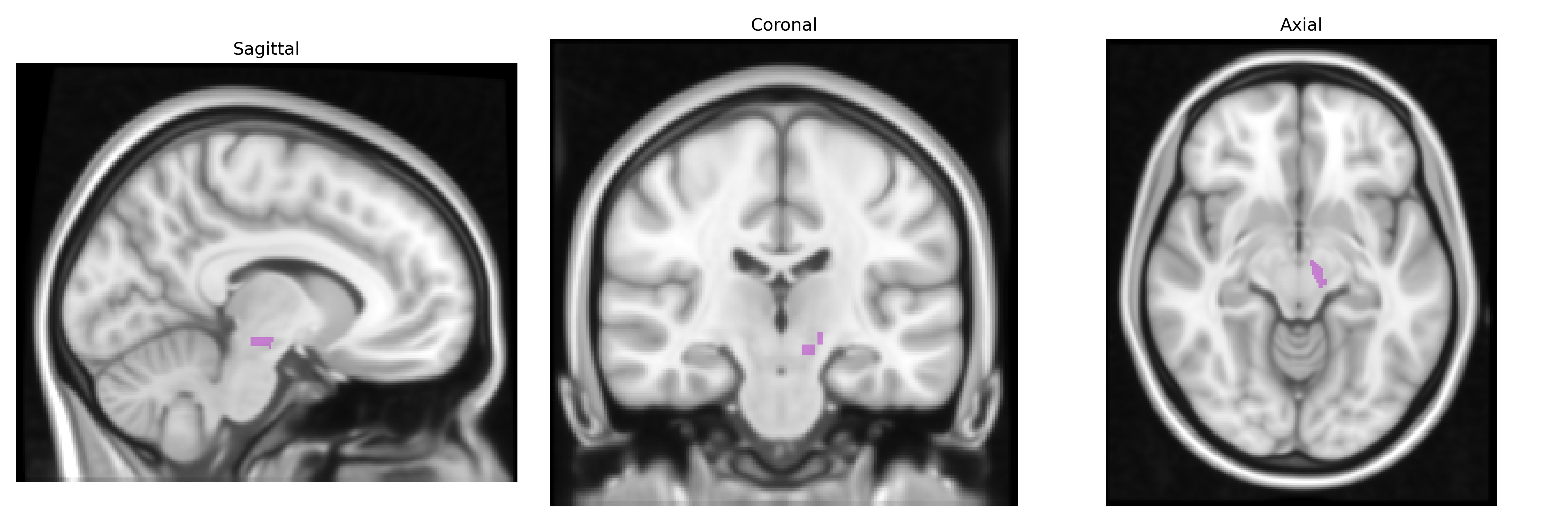
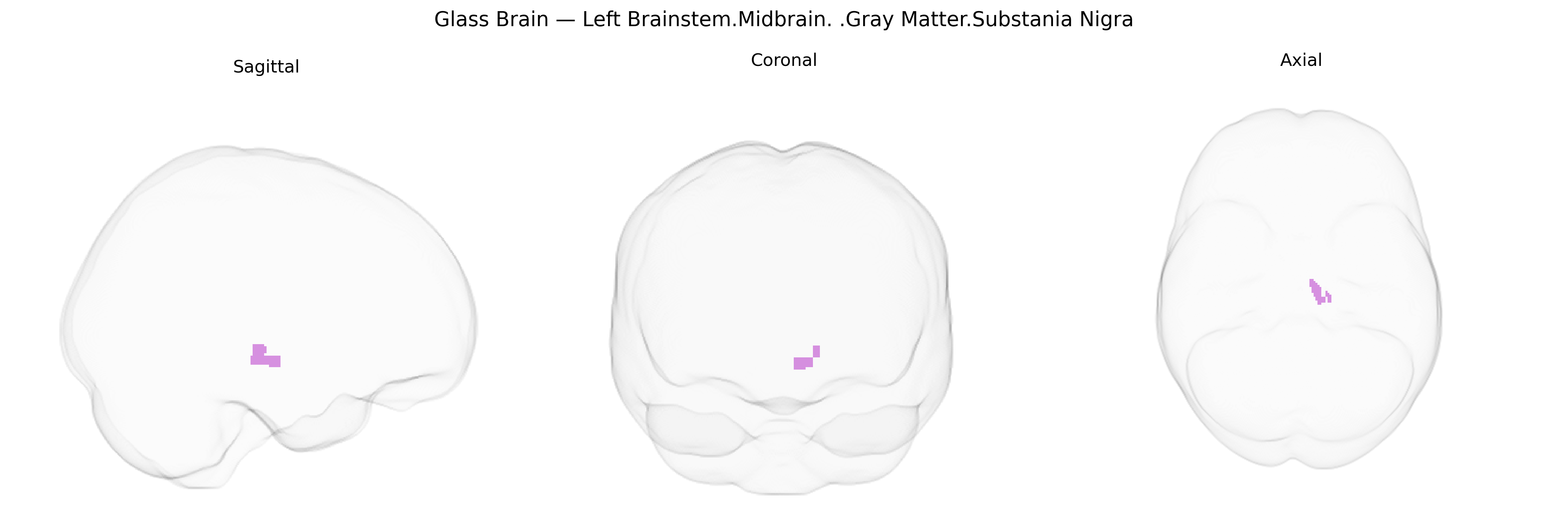
The bilateral Left Brainstem.Midbrain.Gray Matter.Substantia Nigra is a paired, pigmented basal midbrain structure located in the ventral mesencephalon, forming part of the brainstem and classically divided into the pars compacta and pars reticulata. It contains densely packed dopaminergic neurons that project prominently via the nigrostriatal pathway to the dorsal striatum, critically modulating motor control, action selection, and habit formation, as well as contributing to reward and learning circuits. Degeneration of dopaminergic neurons in the substantia nigra pars compacta is a hallmark of Parkinson’s disease, leading to bradykinesia, rigidity, and resting tremor, while dysfunction in its circuitry is also implicated in other movement disorders and some psychiatric conditions. Substantia nigra
The bilateral left brainstem midbrain gray matter substantia nigra—as defined in the Talairach labels 1 mm atlas—is a dopaminergic hub whose structure and function show robust genetic influences and numerous disease associations in GWAS and related genetic studies. Variants in genes critically involved in dopaminergic neuron maintenance and synaptic function, such as SNCA, LRRK2, GBA, MAPT, and PARK2, are strongly associated with Parkinson’s disease, which features hallmark degeneration of substantia nigra pars compacta neurons; these same loci often show pleiotropic effects on related phenotypes such as essential tremor, REM sleep behavior disorder, and dementia with Lewy bodies. Polygenic risk scores for Parkinson’s disease correlate with changes in nigral volume, iron content, and neuromelanin-sensitive MRI signal in imaging–genetics studies, and GWAS of nigral iron and microstructure highlight associations near genes involved in mitochondrial function, iron handling (e.g., HFE), and oxidative stress. Beyond movement disorders, genetic variation influencing midbrain dopaminergic pathways—including polymorphisms in DRD2, DRD3, COMT, and DAT1 (SLC6A3)—has been linked to schizophrenia, bipolar disorder, major depression, ADHD, and addiction-related traits, often via imaging–genetics findings showing altered substantia nigra connectivity or dopaminergic signaling. Structural and functional MRI GWAS using large datasets (e.g., UK Biobank) further suggest that nigral volume and midbrain connectivity are heritable traits associated with common variants in neurodevelopmental and axon-guidance genes, though these effects are typically modest and distributed across many loci.
Overview generated by GPT-4o (2026).
Region ID: 341
Hemisphere: bilateral
Atlas: Talairach labels 1mm
Full Quality Version: Download MP4
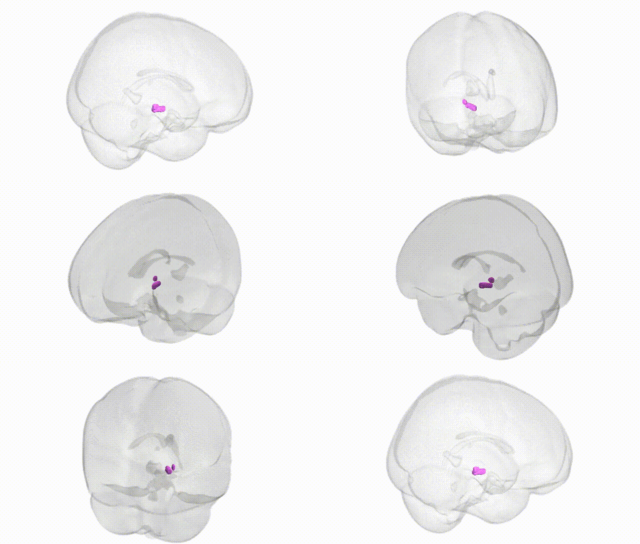
Full Quality Version: Download MP4
Wali Sidiqyar*, Gaurav Rudravaram*, Elyssa M. McMaster, Trent M. Schwartz, Adam M. Saunders, Kurt G. Schilling, Bennett A. Landman "Introducing SPINS: A Shared Public Visualization Library of Neuroanatomical Structures." Medical Imaging with Deep Learning- short paper
This resource is licensed under CC0 1.0 Universal (Public Domain).