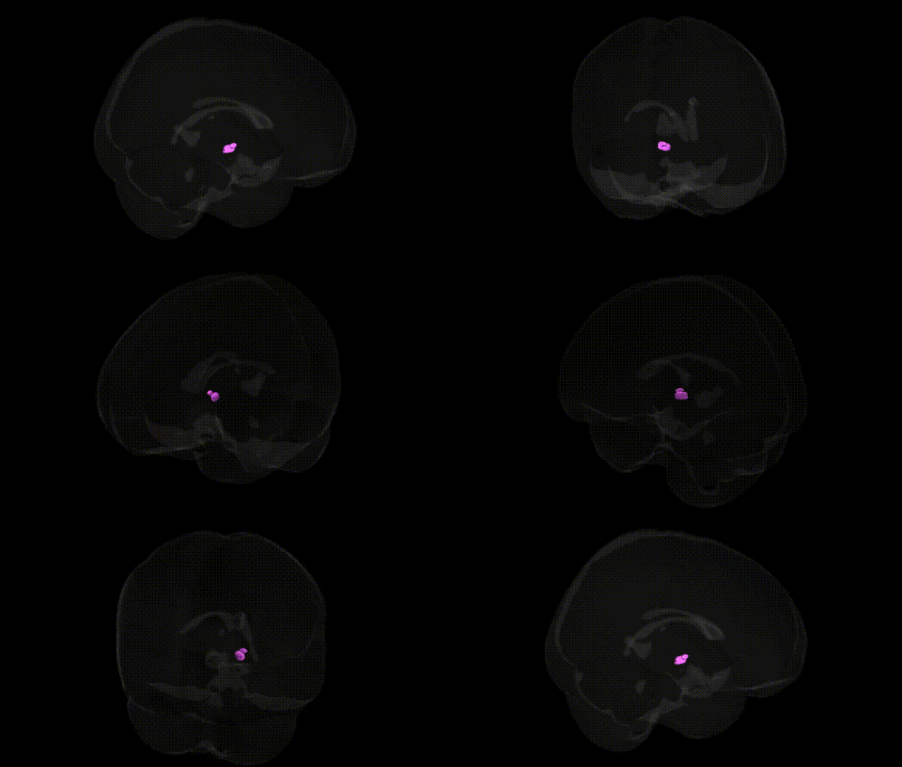
The bilateral Left Brainstem.Midbrain.Gray Matter.Subthalamic Nucleus corresponds to the subthalamic nucleus, a small, lens-shaped gray-matter structure located in the ventral part of the diencephalon, just inferior to the thalamus and medial to the internal capsule, functionally associated with the midbrain and basal ganglia circuitry. It receives excitatory glutamatergic input primarily from the cortex (via the hyperdirect pathway) and inhibitory GABAergic input from the external segment of the globus pallidus, and sends excitatory projections to the internal globus pallidus and substantia nigra pars reticulata. The subthalamic nucleus plays a critical role in the indirect and hyperdirect pathways of motor control, contributing to the suppression of unwanted movements and modulation of motor output; lesions or dysfunction in this nucleus are implicated in movement disorders such as hemiballismus and Parkinson’s disease, and it is a frequent target for deep brain stimulation therapies. Subthalamic nucleus
The subthalamic nucleus (STN), a gray matter structure within the midbrain region of the left brainstem, has been strongly implicated in genetic studies of movement and neuropsychiatric disorders, albeit usually at the circuit rather than single-region level. In Parkinson’s disease, rare high-penetrance variants (e.g., in SNCA, LRRK2, GBA, PRKN) and common GWAS loci influence cortico–basal ganglia–thalamic circuitry in which the STN is a key node, and STN deep brain stimulation outcomes have been examined in relation to variants in genes such as COMT, DRD2, DRD3, and BDNF. GWAS of essential tremor and dystonia also implicate basal ganglia–subthalamic circuitry via risk loci near genes involved in synaptic transmission and cytoskeletal dynamics, though effects are typically distributed across networks. Psychiatric and cognitive GWAS point to STN-related circuits rather than the nucleus in isolation—for example, schizophrenia and bipolar disorder risk loci in glutamatergic, GABAergic, and dopaminergic pathway genes (including DRD2, GRIN2A, CACNA1C, and others) affect fronto–subcortical loops involving the STN, while large imaging-genetics consortia (e.g., ENIGMA) show that polygenic scores for major depressive disorder, ADHD, and schizophrenia correlate with volume and microstructural changes in basal ganglia structures that encompass or interface with the STN. Additionally, variants in genes regulating neurodevelopment (e.g., CNTNAP2, NRG1, and SHANK family genes) and motor control (e.g., KCNMA1, ADCY5) have been linked to phenotypes such as tics, chorea, or hyperkinesias that are modeled through altered STN output. Overall, genetic associations implicate the left subthalamic nucleus as part of broader basal ganglia and fronto–subcortical networks involved in motor, cognitive, and affective traits, but current GWAS generally lack the spatial resolution to ascribe effects exclusively to this specific Talairach-defined region.
Overview generated by GPT-4o (2026).
Region ID: 453
Hemisphere: bilateral
Atlas: Talairach labels 1mm
Full Quality Version: Download MP4
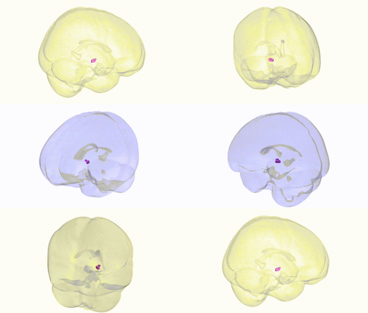
Full Quality Version: Download MP4
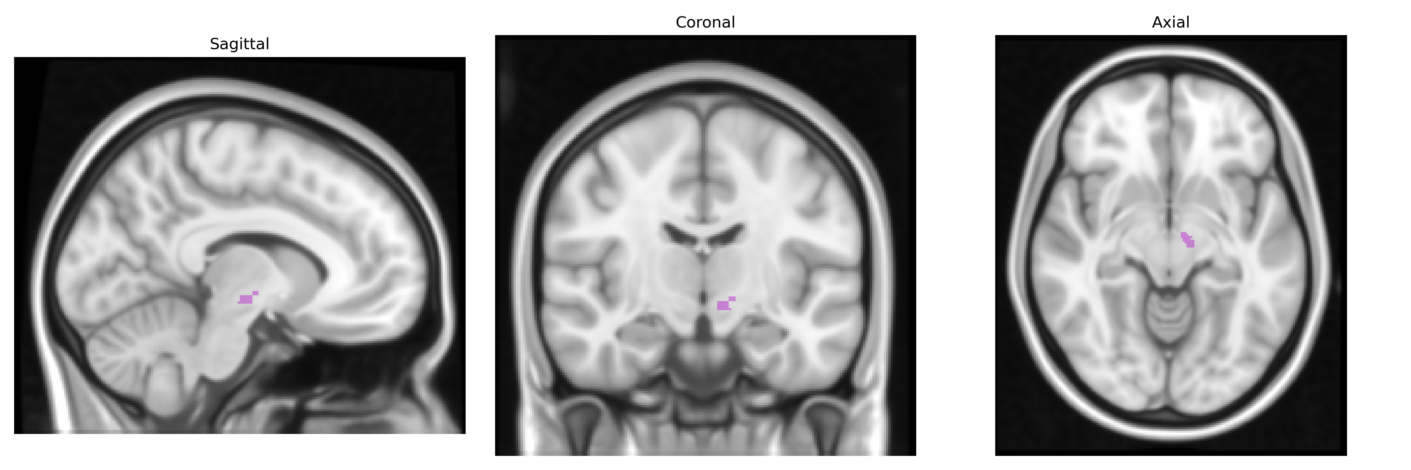
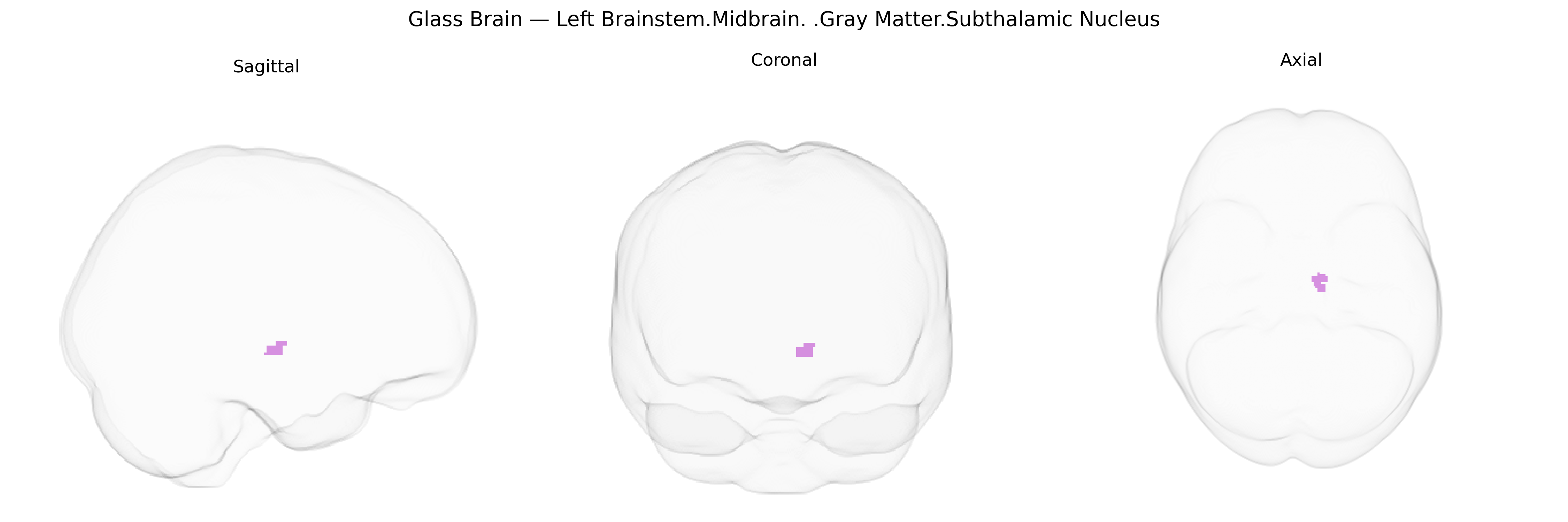
Wali Sidiqyar*, Gaurav Rudravaram*, Elyssa M. McMaster, Trent M. Schwartz, Adam M. Saunders, Kurt G. Schilling, Bennett A. Landman "Introducing SPINS: A Shared Public Visualization Library of Neuroanatomical Structures." Medical Imaging with Deep Learning- short paper
This resource is licensed under CC0 1.0 Universal (Public Domain).