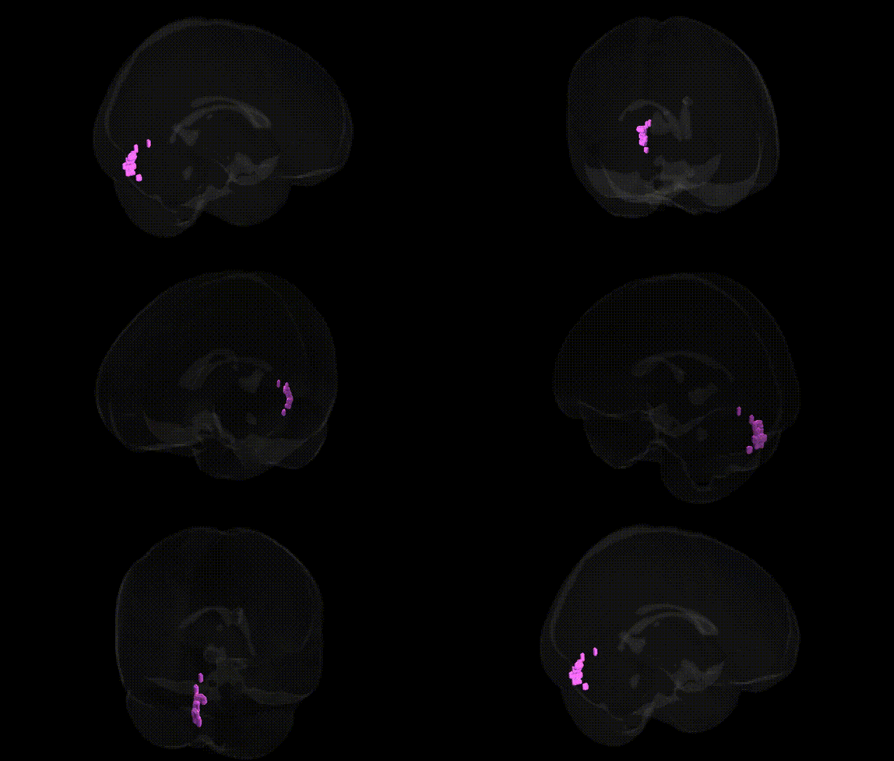
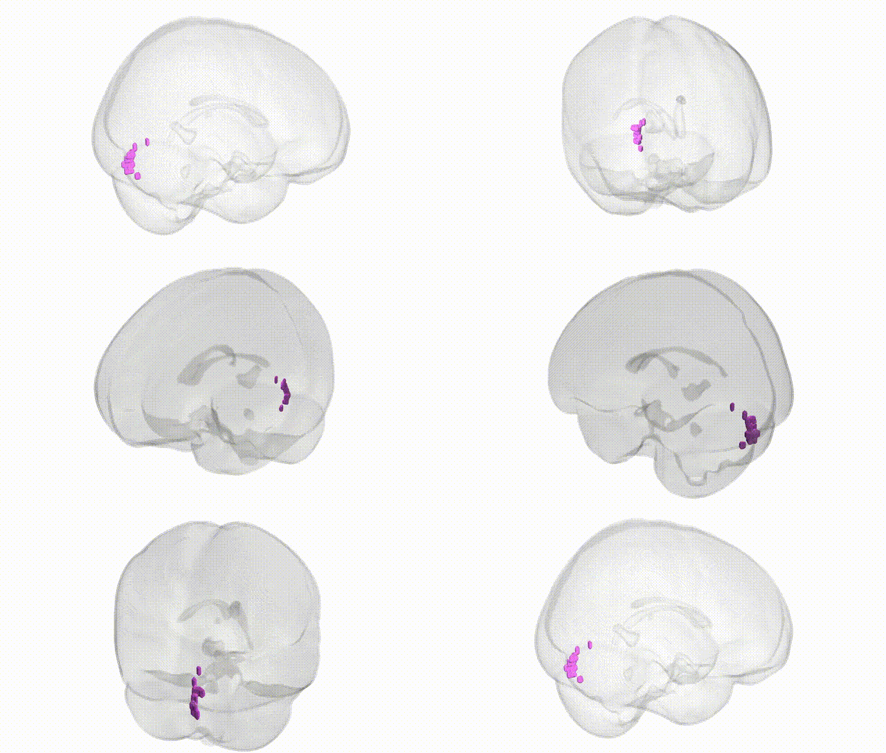
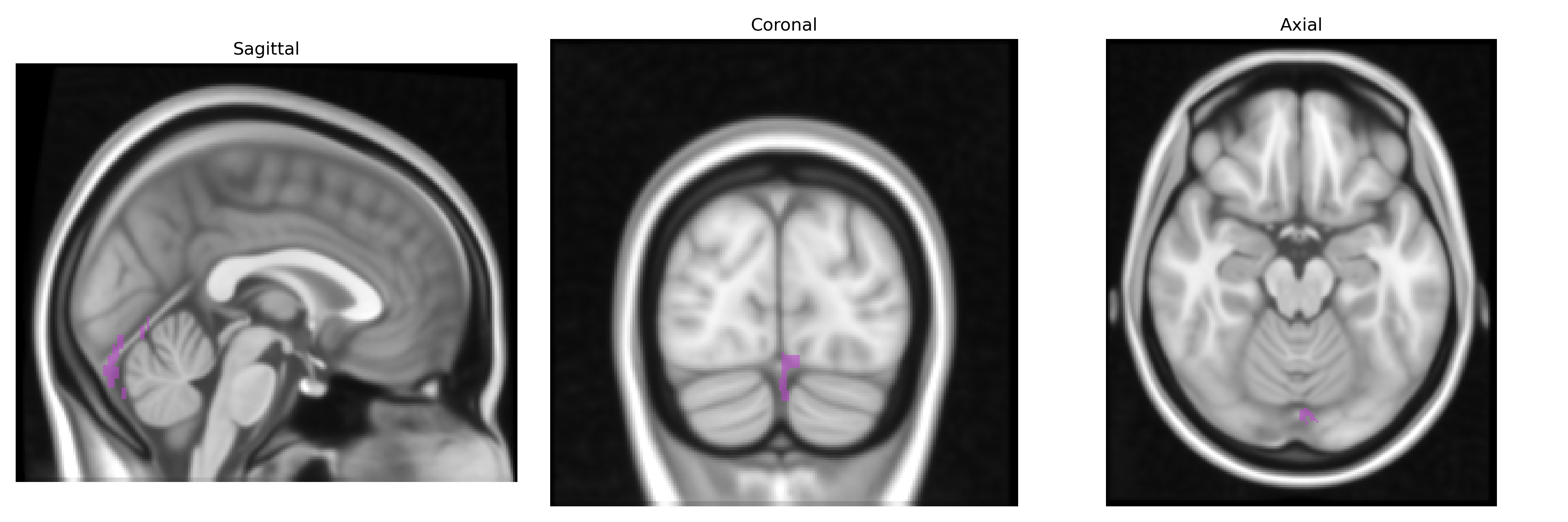
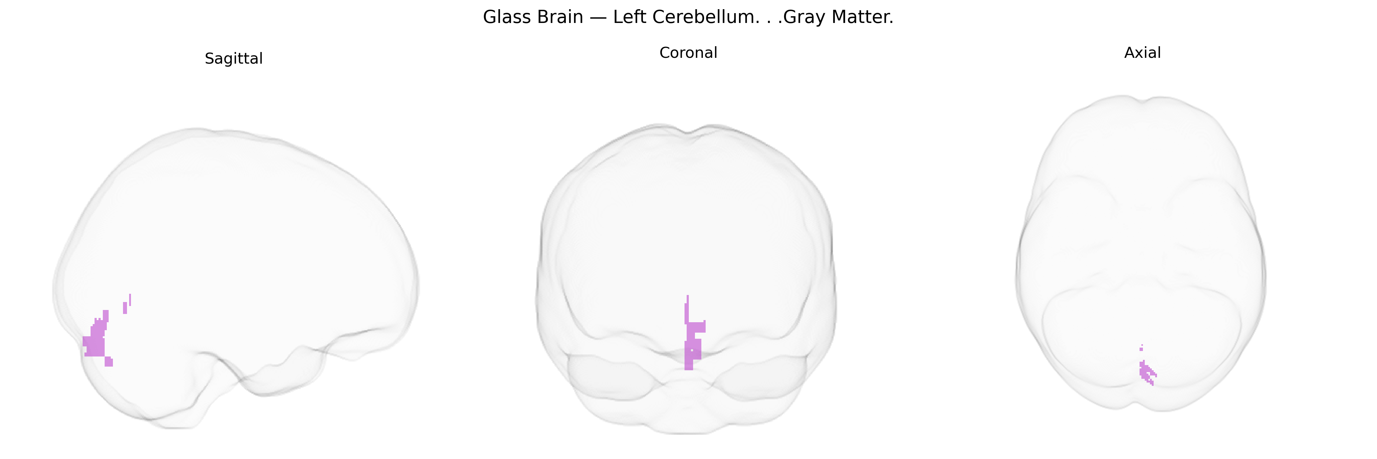
The bilateral Left Cerebellum gray matter comprises the cortical neuronal layers of the cerebellar hemisphere on the left side, which is involved in coordination of voluntary movements, maintenance of posture and balance, and fine-tuning of motor commands via its extensive connections with the cerebral cortex, brainstem, and spinal cord. This region contributes to error correction in motor execution, motor learning (including adaptation and timing of movements), and also participates in certain cognitive and affective processes through its connections with association cortices and limbic structures. Architecturally, it is organized into folia containing a highly regular laminar circuitry of Purkinje cells, granule cells, and interneurons that integrate mossy and climbing fiber inputs to modulate deep cerebellar nuclei output. Cerebellum
The left cerebellar gray matter has been repeatedly implicated in genetic and GWAS findings related to motor coordination, cognition, and neuropsychiatric vulnerability, although most large-scale studies analyze total cerebellar or lobule-specific volumes rather than Talairach-defined parcels. Twin and heritability studies show moderate-to-high genetic influence on cerebellar gray matter volume (often h² ~0.4–0.7), and multivariate GWAS of brain structure (e.g., ENIGMA, UK Biobank) have identified common variants near genes involved in neurodevelopment, synaptic function, and axon guidance (such as MAPT, PLP1, and genes in Wnt and Notch pathways) associated with cerebellar morphology. Polygenic overlap has been reported between cerebellar volume and cognitive ability, educational attainment, and neurodevelopmental disorders, with cerebellar gray matter consistently implicated in schizophrenia, autism spectrum disorder, major depression, and ADHD through imaging–genetics and imaging–GWAS approaches that show altered cerebellar structure or connectivity linked to disorder-associated polygenic risk scores. Additionally, rare variant and CNV studies in disorders with strong cerebellar phenotypes (e.g., spinocerebellar ataxias, 22q11.2 deletion syndrome) underline the sensitivity of left cerebellar gray matter to disruptions in genes governing neuronal migration and synaptic plasticity, supporting a genetically mediated role for this region in both motor and higher-order cognitive and affective functions.
Overview generated by GPT-4o (2026).
Region ID: 66
Hemisphere: bilateral
Atlas: Talairach labels 1mm
Full Quality Version: Download MP4
Full Quality Version: Download MP4
Wali Sidiqyar*, Gaurav Rudravaram*, Elyssa M. McMaster, Trent M. Schwartz, Adam M. Saunders, Kurt G. Schilling, Bennett A. Landman "Introducing SPINS: A Shared Public Visualization Library of Neuroanatomical Structures." Medical Imaging with Deep Learning- short paper
This resource is licensed under CC0 1.0 Universal (Public Domain).