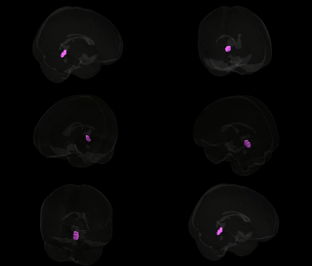
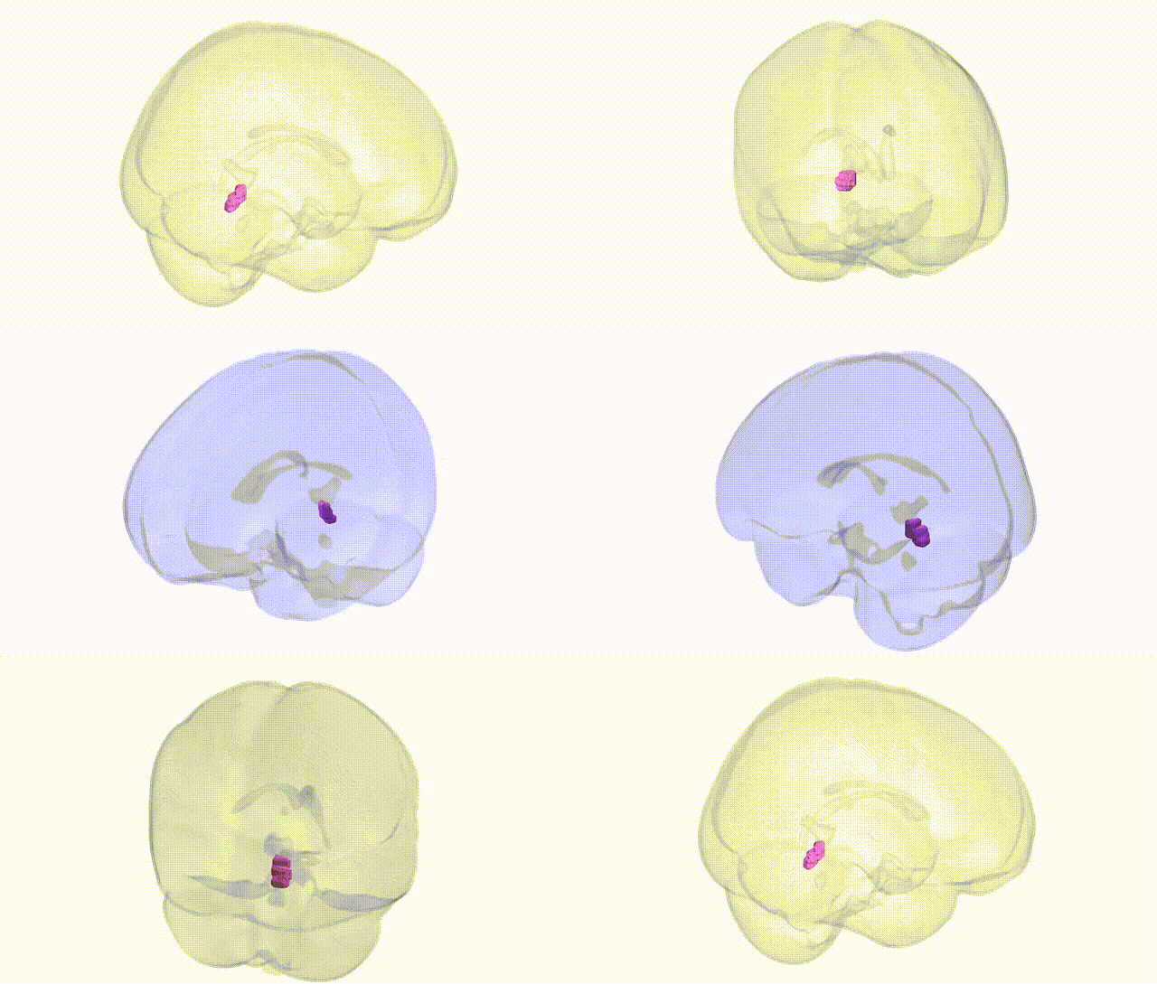
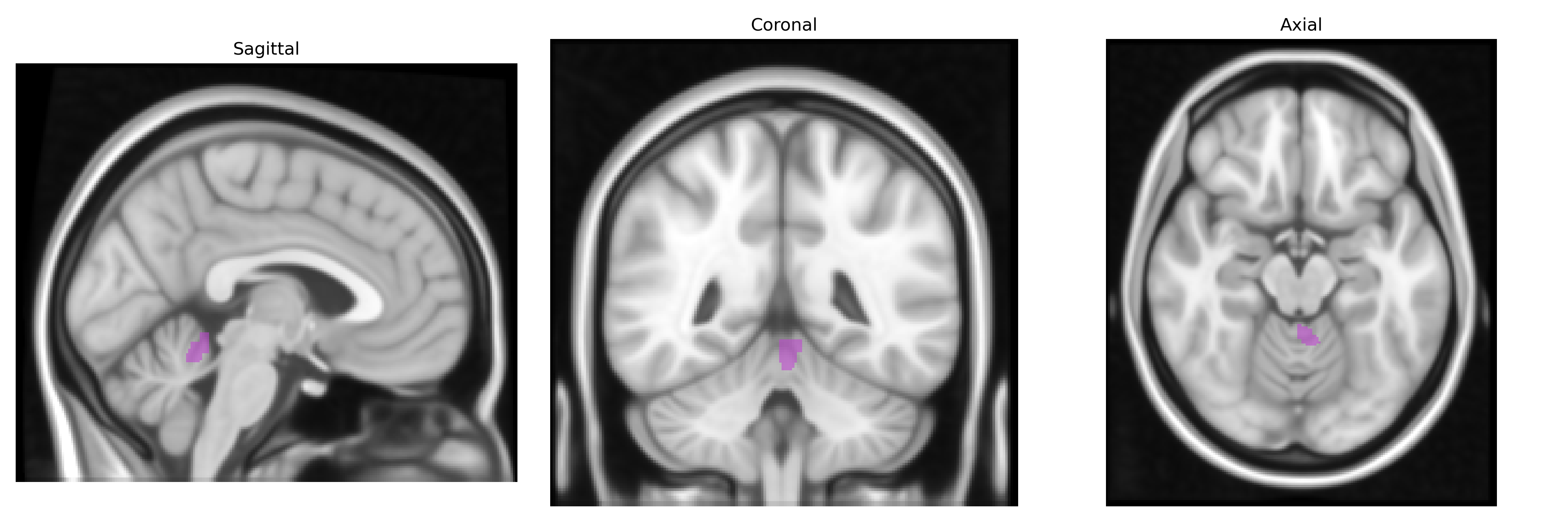
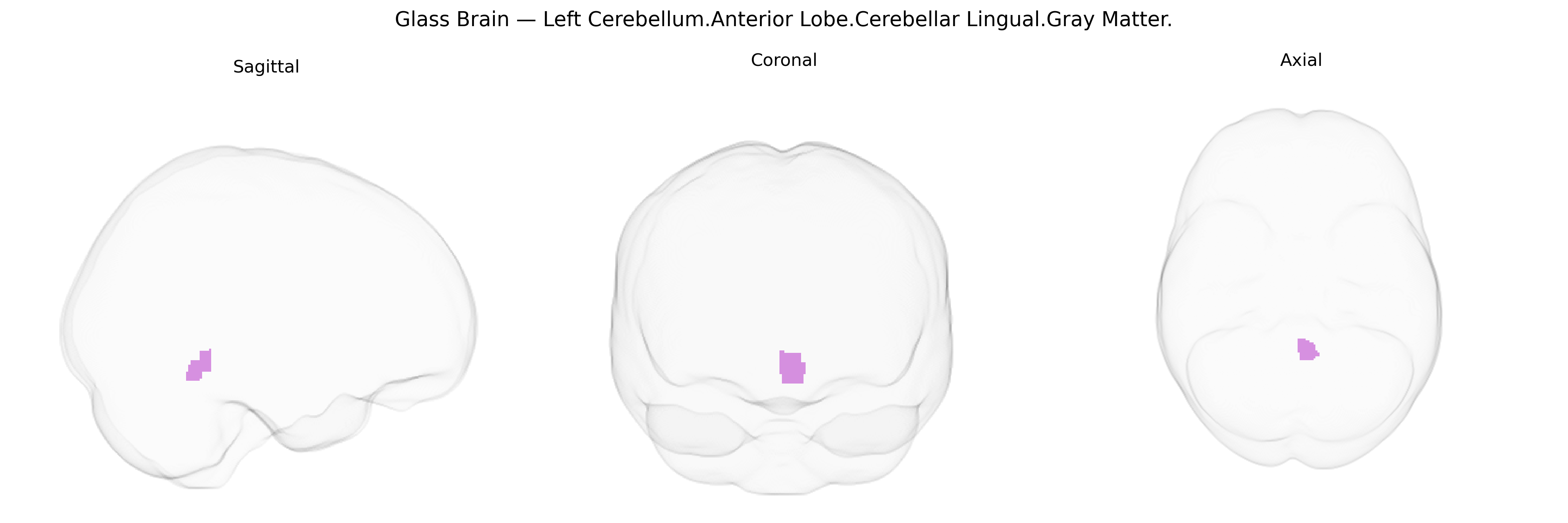
The bilateral Left Cerebellum.Anterior Lobe.Cerebellar Lingual.Gray Matter refers to gray matter within the lingula (also called the lingual lobule) of the anterior lobe of the cerebellum, located medially along the superior surface adjacent to the vermis. This region comprises cortical neurons involved in fine-tuning motor activity, particularly in the regulation of posture, balance, and coordination of limb movements via cerebellar circuits that integrate proprioceptive and vestibular inputs. As part of the anterior lobe, the lingula contributes to the modulation of ongoing movements and motor learning, influencing timing and precision of motor output through its connections with deep cerebellar nuclei and descending motor pathways. There is no direct link; related structure: Cerebellum.
The bilateral Left Cerebellum.Anterior Lobe.Cerebellar Lingual.Gray Matter region, as defined in the Talairach 1 mm atlas, lies within anterior cerebellar territories implicated in motor coordination, oculomotor control, and cognitive timing, and is often mapped within broader cerebellar lobules I–V and adjacent lobule VI in imaging-genetics work rather than as a distinct parcel. Large-scale GWAS of brain morphology (e.g., ENIGMA, UK Biobank) have identified multiple loci (including variants near or within genes such as KCNK2, RELN, MAPT, and genes involved in neurodevelopment, synaptic function, and axon guidance) associated with global and lobule-specific cerebellar gray matter volume, though these studies typically report effects at the level of whole anterior cerebellum or specific lobules rather than the lingual subregion per se. Polygenic architectures underlying cerebellar volume overlap with genetic risk for neurodevelopmental and psychiatric disorders—including schizophrenia, major depressive disorder, bipolar disorder, autism spectrum disorder, and ADHD—as well as cognitive traits such as general intelligence and educational attainment, consistent with well-documented cerebellar involvement in cortico-cerebellar circuits. Cerebellar structural variation encompassing this region has also been linked, in imaging-genetic and candidate-gene studies, to spinocerebellar ataxias, dystonia, essential tremor, and some epilepsies, though these typically implicate widespread cerebellar networks rather than isolating this specific anterior lingual zone. Overall, current genetic evidence points to shared polygenic influences on cerebellar gray matter in this area and a range of motor, cognitive, and psychiatric phenotypes, but no robust GWAS has yet isolated unique, region-specific genetic associations restricted to the Cerebellar Lingual.Gray Matter label itself.
Overview generated by GPT-4o (2026).
Region ID: 211
Hemisphere: bilateral
Atlas: Talairach labels 1mm
Full Quality Version: Download MP4
Full Quality Version: Download MP4
Wali Sidiqyar*, Gaurav Rudravaram*, Elyssa M. McMaster, Trent M. Schwartz, Adam M. Saunders, Kurt G. Schilling, Bennett A. Landman "Introducing SPINS: A Shared Public Visualization Library of Neuroanatomical Structures." Medical Imaging with Deep Learning- short paper
This resource is licensed under CC0 1.0 Universal (Public Domain).