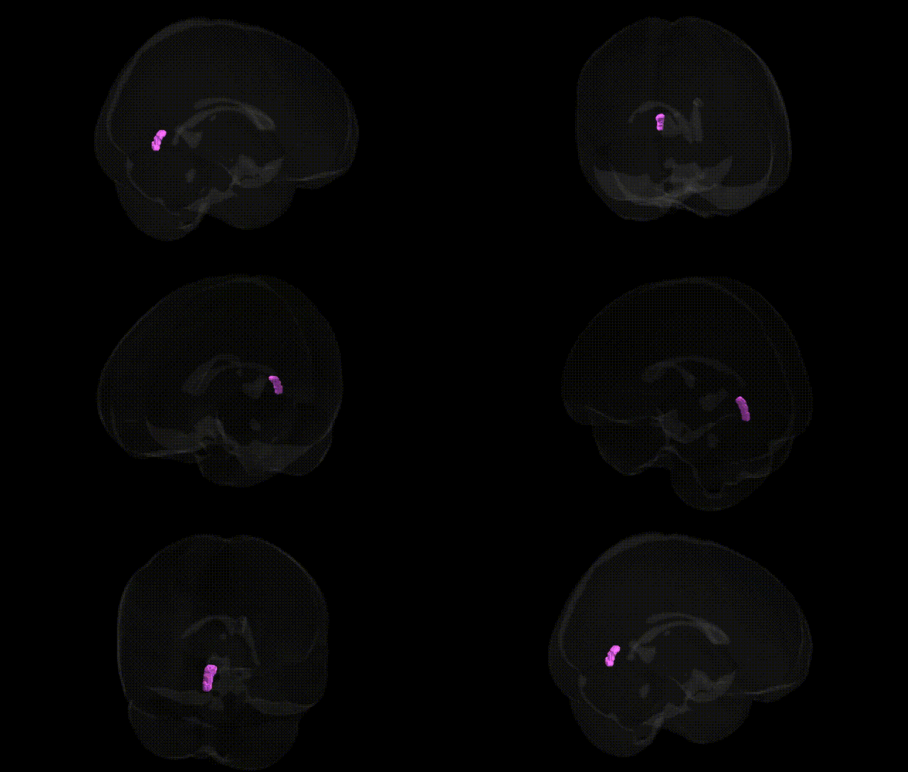
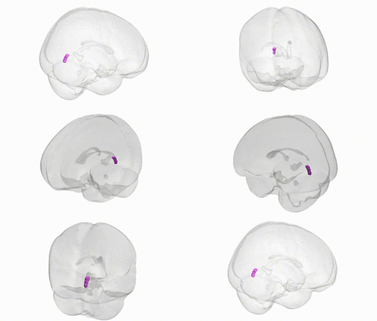
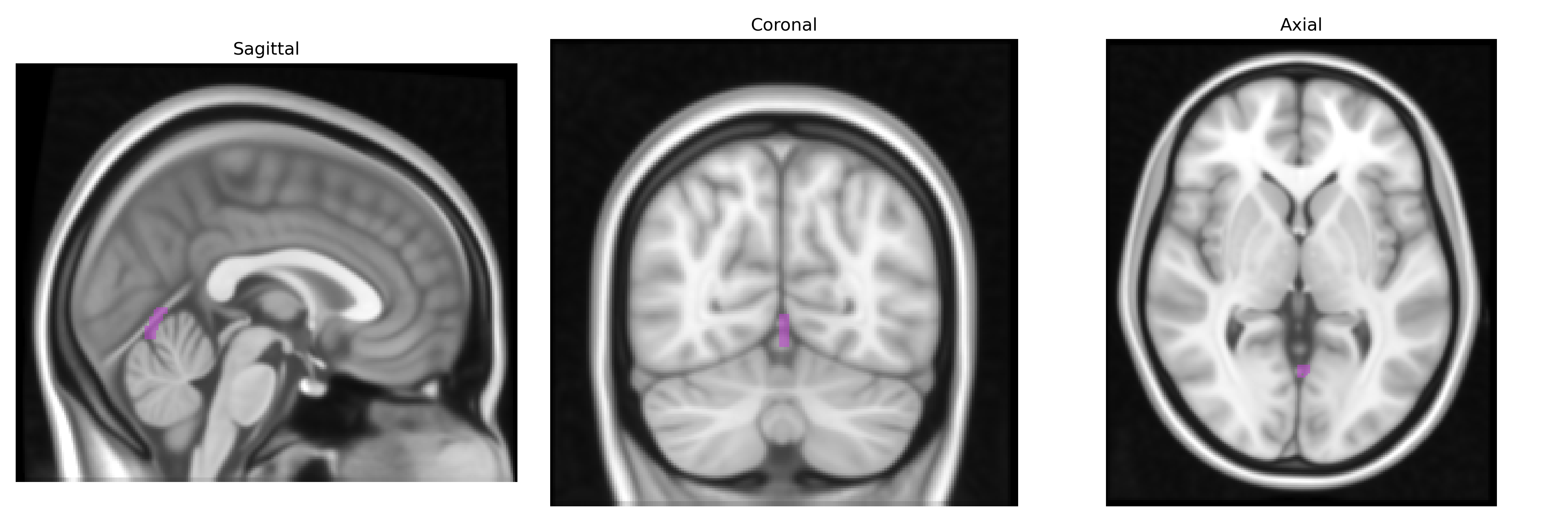
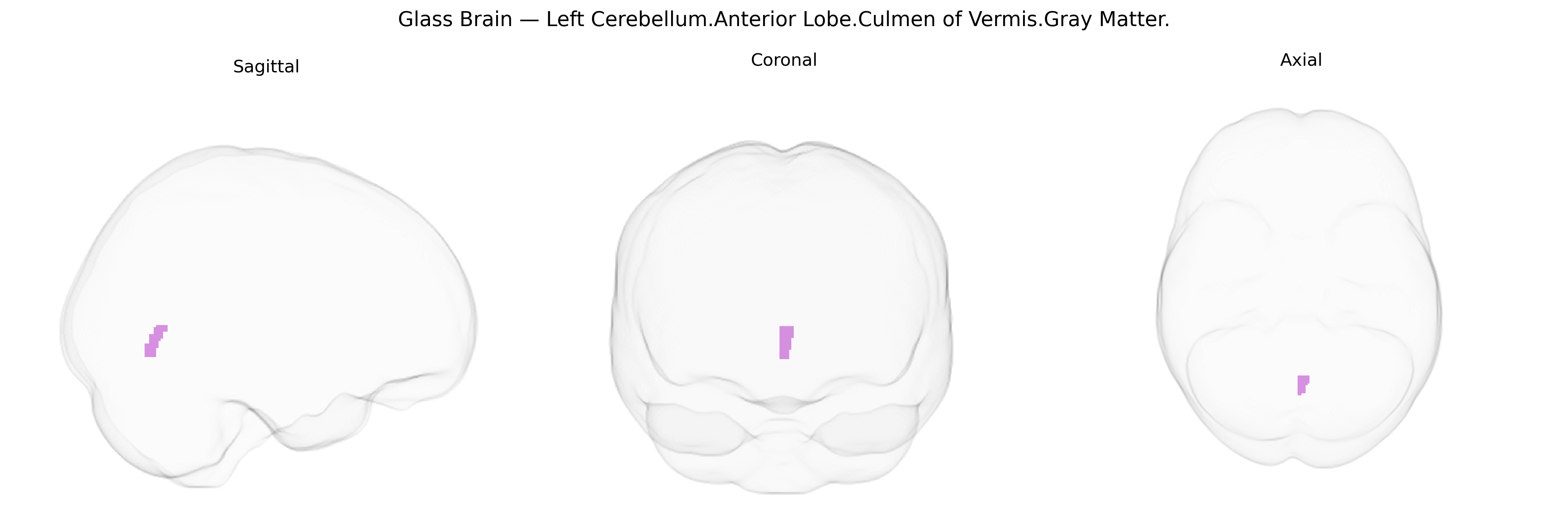
The bilateral Left Cerebellum.Anterior Lobe.Culmen of Vermis.Gray Matter corresponds to gray matter within the culmen, a midline (vermal) structure in the anterior lobe of the cerebellum that participates in motor coordination, posture, and muscle tone regulation. As part of the spinocerebellum, this region receives substantial proprioceptive input from the spinal cord and integrates this information to fine-tune axial and proximal limb movements, contributing to balance and gait. Neuronal circuitry in the culmen (including Purkinje cells and deep cerebellar nuclei projections) modulates descending motor pathways, and lesions in this area are associated with truncal ataxia, dysmetria, and impaired postural control. There is no specific Wikipedia article for the culmen of the vermis, but it is a subregion of the cerebellar vermis: Cerebellar vermis.
The bilateral left cerebellar anterior lobe culmen of the vermis (gray matter) occupies a region broadly implicated in motor coordination, postural control, and aspects of cognitive and affective processing, and genetic associations are typically reported at the level of the anterior cerebellar vermis or lobules IV–V rather than this exact Talairach-defined parcel. Large-scale imaging–genetics consortia (e.g., ENIGMA, UK Biobank) have shown that anterior cerebellar gray-matter volume and morphology are highly heritable and influenced by numerous common variants of small effect, with polygenic architectures overlapping those for general brain size, height, and intracranial volume, but no single variant or locus is uniquely and reproducibly assigned to this specific culmen-of-vermis label. GWAS of cerebellar volume and surface area have identified loci near genes involved in neurodevelopment and synaptic function (such as variants in or near KIAA0586, PAPPA2, and other neurodevelopmental genes), and these often show pleiotropic effects on psychiatric or cognitive phenotypes, though regional specificity seldom resolves to the culmen vermis alone. Disorders in which anterior vermis abnormalities are repeatedly noted—often with a genetic component—include alcohol use disorder (with vermian atrophy associated with variants affecting glutamatergic and GABAergic signaling), autism spectrum disorder and other neurodevelopmental conditions (in which rare de novo and inherited variants in genes regulating cerebellar development, e.g., in synaptic and chromatin-remodeling pathways, are implicated), and some spinocerebellar ataxias caused by repeat expansions (e.g., in ATXN genes) that produce prominent anterior vermis degeneration. Additional associations arise in mood and psychotic disorders through imaging–genetics studies linking polygenic risk scores for major depression, bipolar disorder, and schizophrenia with structural or functional changes in midline cerebellar regions including the culmen of the vermis, though these effects are modest and not region-specific; overall, current evidence supports a broadly polygenic, pleiotropic influence on this region’s structure and function rather than discrete, well-validated locus-level associations unique to this Talairach-defined area.
Overview generated by GPT-4o (2026).
Region ID: 321
Hemisphere: bilateral
Atlas: Talairach labels 1mm
Full Quality Version: Download MP4
Full Quality Version: Download MP4
Wali Sidiqyar*, Gaurav Rudravaram*, Elyssa M. McMaster, Trent M. Schwartz, Adam M. Saunders, Kurt G. Schilling, Bennett A. Landman "Introducing SPINS: A Shared Public Visualization Library of Neuroanatomical Structures." Medical Imaging with Deep Learning- short paper
This resource is licensed under CC0 1.0 Universal (Public Domain).