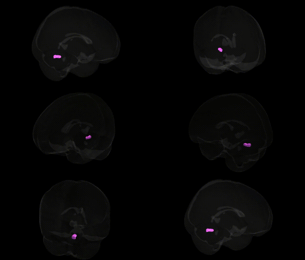
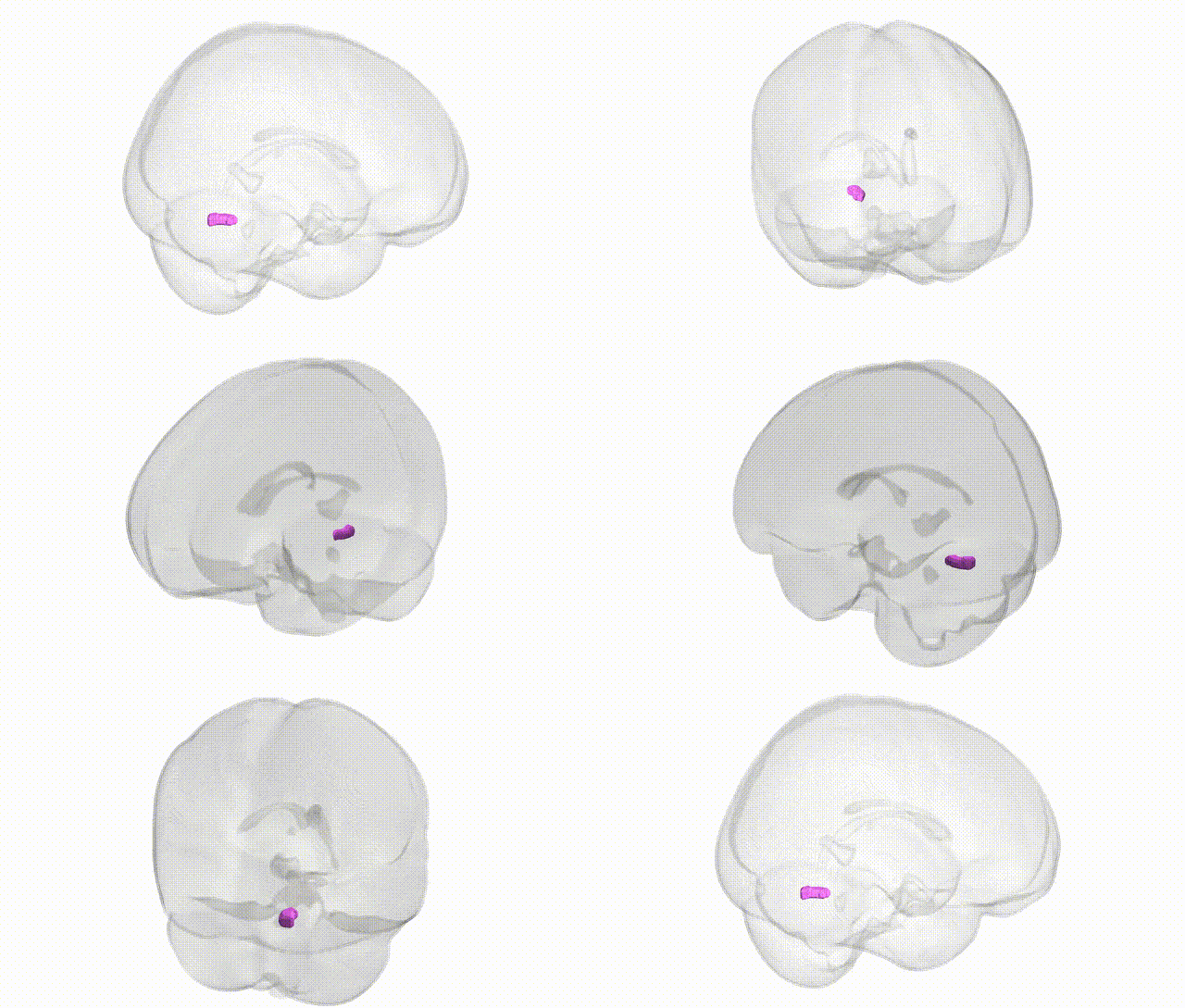
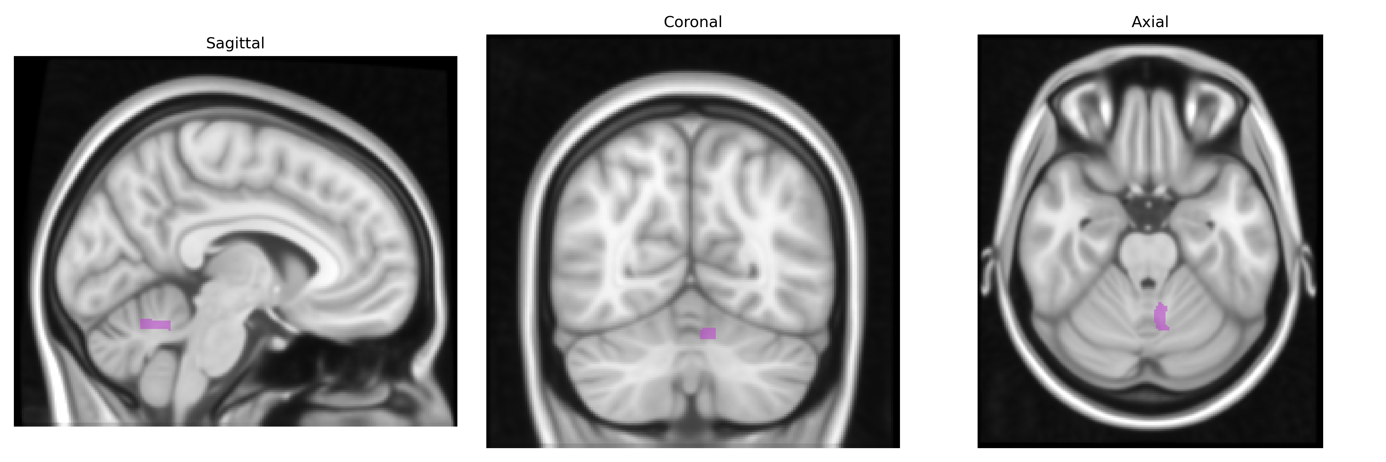
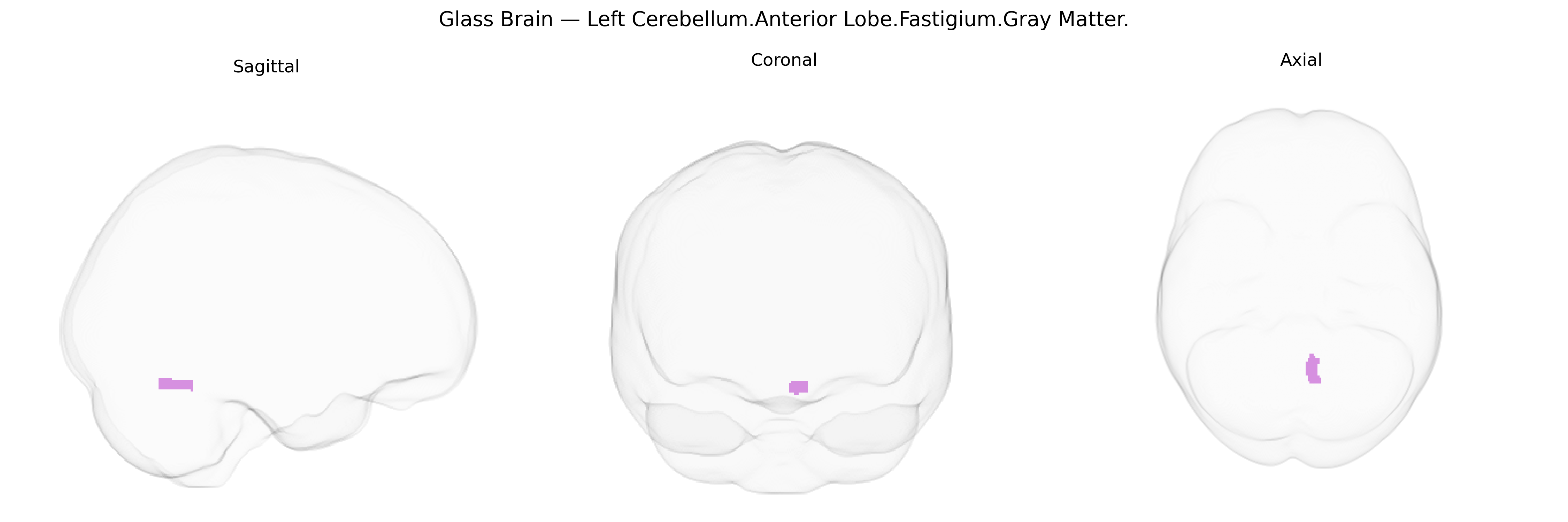
The bilateral Left Cerebellum, Anterior Lobe, Fastigium, Gray Matter corresponds to midline cerebellar gray matter within the anterior lobe, centered around the fastigial nucleus and adjacent vermal regions. This region is involved in posture, balance, and the coordination of proximal and axial musculature, integrating somatosensory and vestibular inputs to modulate motor output via cerebellar and brainstem pathways. Functionally, the fastigial region contributes to the regulation of muscle tone, stance, and locomotion, and participates in the control of eye and head movements through its connections with vestibular and reticular nuclei. In the Talairach 1 mm atlas, this label reflects gray matter in the anterior cerebellar vermis and associated deep nuclei, forming part of the spinocerebellar circuitry critical for real-time error correction during movement. There is no direct link for this exact composite label; related structure: Cerebellum.
The bilateral Left Cerebellum.Anterior Lobe.Fastigium.Gray Matter region, a midline cerebellar structure involved in posture, balance, and affective regulation, has been implicated indirectly in several genetic and genome-wide association findings, though few studies target this subregion with high anatomical specificity. Imaging–genetics analyses and cerebellar volume GWAS indicate that common variants in genes related to neurodevelopment (e.g., MAPT, FOXP2, and synaptic or axonal guidance genes) and general brain structure (e.g., loci near KTN1 and genes influencing intracranial volume) contribute to inter-individual variation in cerebellar gray matter, including anterior lobe and vermal areas encompassing the fastigial region. Polygenic risk for psychiatric and neurodevelopmental disorders—such as schizophrenia, major depressive disorder, bipolar disorder, and autism spectrum disorder—has been associated with altered cerebellar vermis and anterior lobe morphology or activation, suggesting shared genetic architecture between these conditions and fastigial-region structure or function. Rare-variant and familial studies in spinocerebellar ataxias, dystonia, and congenital cerebellar malformations also implicate genes controlling Purkinje cell integrity, ion channel function, and cerebellar patterning (such as CACNA1A, ATXN genes, and homeobox/segmental patterning genes), which affect midline cerebellar nuclei including the fastigial nucleus. However, current large-scale GWAS catalog entries and neuroimaging consortia reports typically reference broader cerebellar lobular or vermal volumes, so evidence for genetic associations specific to the Talairach-defined Left Cerebellum.Anterior Lobe.Fastigium.Gray Matter region remains indirect and largely inferred from studies of overall cerebellar structure, anterior lobe measures, or vermis-focused analyses.
Overview generated by GPT-4o (2026).
Region ID: 165
Hemisphere: bilateral
Atlas: Talairach labels 1mm
Full Quality Version: Download MP4
Full Quality Version: Download MP4
Wali Sidiqyar*, Gaurav Rudravaram*, Elyssa M. McMaster, Trent M. Schwartz, Adam M. Saunders, Kurt G. Schilling, Bennett A. Landman "Introducing SPINS: A Shared Public Visualization Library of Neuroanatomical Structures." Medical Imaging with Deep Learning- short paper
This resource is licensed under CC0 1.0 Universal (Public Domain).