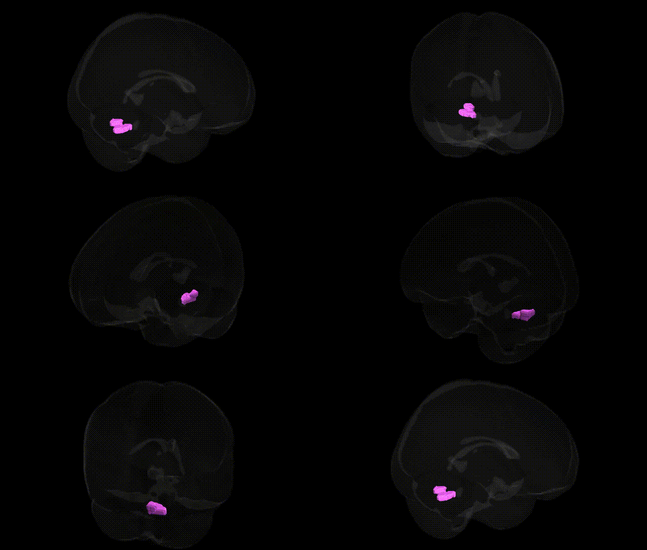
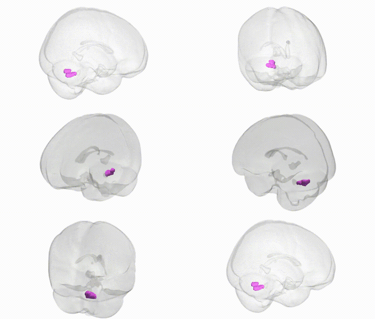
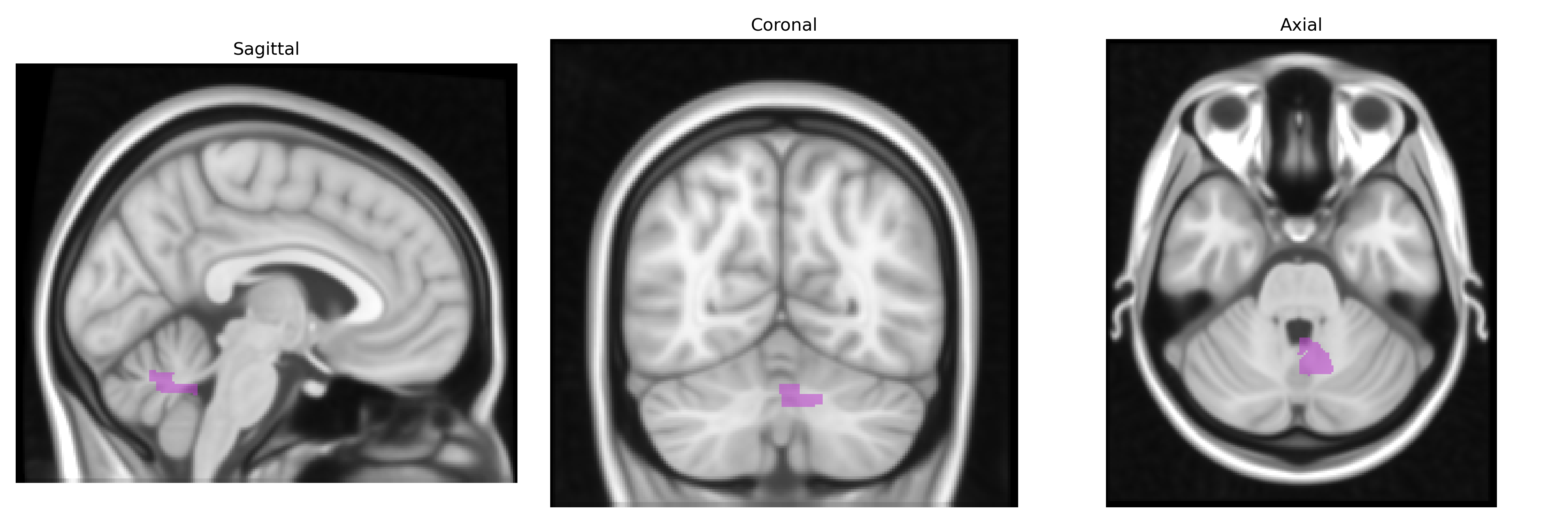
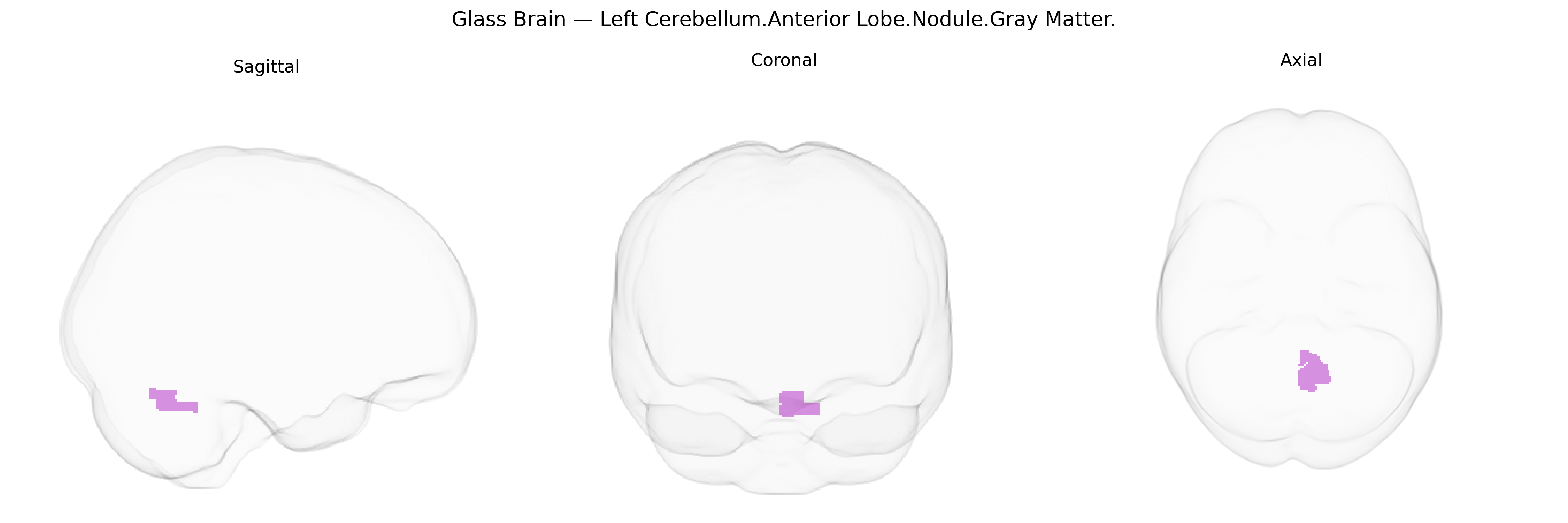
The bilateral Left Cerebellum.Anterior Lobe.Nodule.Gray Matter corresponds to gray matter within the cerebellar nodule, the most medial component of the anterior lobe and a central element of the flocculonodular (vestibulocerebellum) region. This structure receives dense vestibular and proprioceptive input and plays a critical role in balance, eye movement control, and coordination of axial and proximal limb musculature. Neuronal circuitry in the nodule integrates sensory information to fine-tune postural reflexes and maintain equilibrium, working closely with vestibular nuclei and other cerebellar lobules. Lesions involving the nodule can lead to truncal ataxia, gait disturbances, and impaired vestibulo-ocular reflexes, reflecting its importance in stabilizing the body and eyes in space. There is no direct link for this exact Talairach-labeled region; a closely related structure is the Flocculonodular lobe.
The bilateral left cerebellar anterior lobe nodule (gray matter) has been implicated in several genetic and GWAS-based associations, primarily via large-scale imaging-genetics and psychiatric genetics studies that map SNPs to cerebellar volume, microstructure, or functional connectivity rather than to this small subregion specifically. Variants in genes involved in neurodevelopment, synaptic function, and axonal guidance (for example, MIR924HG, PAX3, and others highlighted in ENIGMA and UK Biobank cerebellar GWAS) have been associated with regional cerebellar morphology that includes the anterior lobe and midline vermis regions encompassing the nodule. Cerebellar midline and anterior lobe structures, including the nodule, have shown altered volume or connectivity in individuals with autism spectrum disorder, schizophrenia, bipolar disorder, ADHD, and major depression, and polygenic risk scores for these disorders correlate with cerebellar measures in imaging-genetics cohorts. Additional GWAS have linked cerebellar volume and function to genes related to GABAergic and glutamatergic signaling, neuronal migration, and calcium channel function, which overlap with risk architectures for neurodevelopmental and psychiatric conditions. However, genetic associations resolved explicitly to the bilateral left anterior lobe nodule in the Talairach 1 mm atlas are sparse, and most evidence derives from broader lobular or vermal cerebellar parcellations, making current genetic findings for this precise nodule-level region indirect and inferred from larger-scale cerebellar phenotypes.
Overview generated by GPT-4o (2026).
Region ID: 84
Hemisphere: bilateral
Atlas: Talairach labels 1mm
Full Quality Version: Download MP4
Full Quality Version: Download MP4
Wali Sidiqyar*, Gaurav Rudravaram*, Elyssa M. McMaster, Trent M. Schwartz, Adam M. Saunders, Kurt G. Schilling, Bennett A. Landman "Introducing SPINS: A Shared Public Visualization Library of Neuroanatomical Structures." Medical Imaging with Deep Learning- short paper
This resource is licensed under CC0 1.0 Universal (Public Domain).