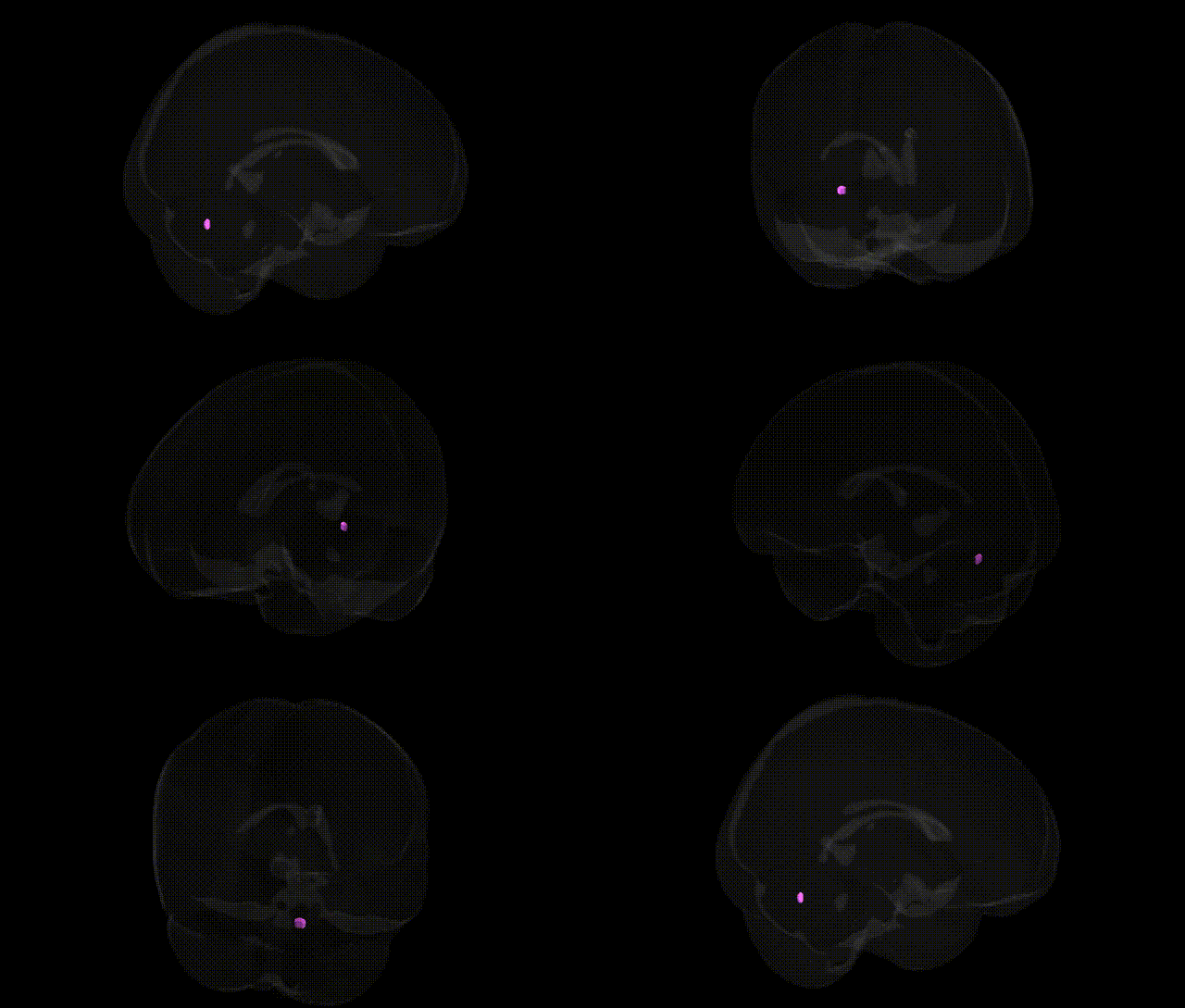
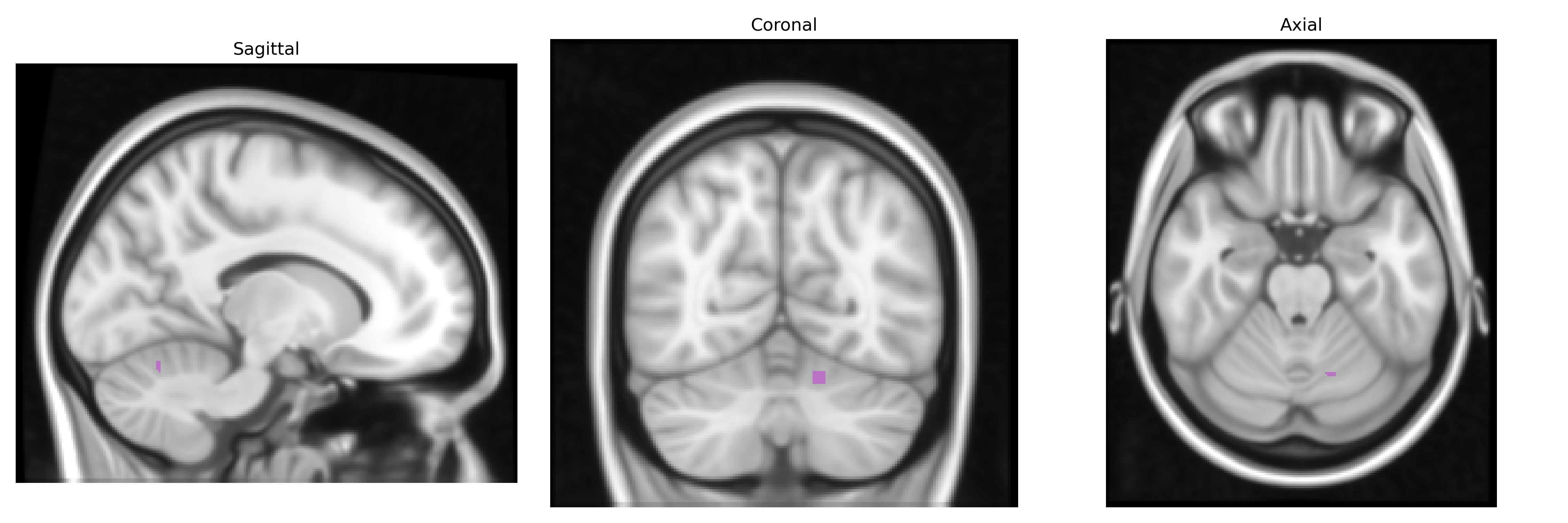
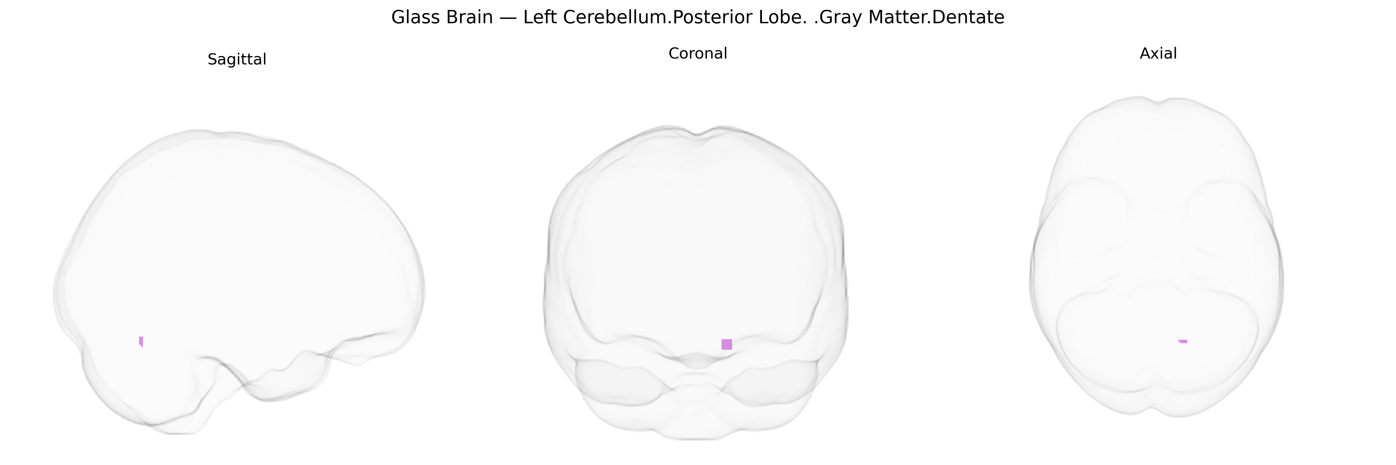
The bilateral Left Cerebellum posterior lobe gray matter of the dentate nucleus is a deep cerebellar structure embedded within the cerebellar white matter and represents the largest and most lateral of the cerebellar nuclei. It receives processed input from the lateral (cerebrocerebellar) hemispheres of the posterior lobe and serves as a major relay station for cerebellar output to motor and premotor cortical areas via the dentatothalamic pathway, thereby contributing to the planning, coordination, and precision of voluntary movements, as well as higher-order cognitive and visuomotor functions. Its organization shows a predominantly somatotopic representation of limb and orofacial musculature, with extensive connections to the contralateral motor cortex and association areas, aligning with its role in fine-tuning complex motor sequences and aspects of motor learning. Dentate nucleus
The bilateral left cerebellum posterior lobe gray matter of the dentate nucleus has been repeatedly implicated in genetic studies of brain structure, cognition, and neuropsychiatric disease, although most findings come from broader cerebellar or lobar measures rather than Talairach-defined 1 mm dentate parcels specifically. Large GWAS of cerebellar volume and morphology (e.g., ENIGMA, UK Biobank–based studies) identify polygenic influences with loci near genes involved in neurodevelopment, synaptic function, and axon guidance (such as FOXP1/FOXP2 regions, MAPT locus on 17q21, and multiple calcium-channel and glutamatergic signaling genes), and show that posterior cerebellar volumes, including dentate-adjacent gray matter, share genetic correlations with general intelligence, educational attainment, and cortical association networks. Genetic overlap has also been reported between cerebellar structure and risk for schizophrenia, major depressive disorder, bipolar disorder, and autism spectrum disorder, consistent with functional MRI evidence that the posterior cerebellum and dentate nucleus participate in cognitive and affective circuits; specific risk loci (e.g., in CACNA1C, GRIN2A, and other synaptic genes) contribute to this shared architecture rather than uniquely targeting the dentate. Additionally, rare Mendelian and copy-number variants affecting cerebellar development (e.g., in genes such as PTF1A, KIAA2022, and some tubulin-related genes) can produce dentate and posterior lobe hypoplasia or atrophy, while spinocerebellar ataxias (notably SCA1, SCA2, SCA3, and SCA6) often show prominent dentate involvement, linking polyglutamine expansion or ion-channel genes to this region. Overall, existing genetic evidence points to a highly polygenic, pleiotropic architecture in which variants influencing neurodevelopmental patterning, synaptic plasticity, and psychiatric risk partially converge on posterior cerebellar and dentate gray matter, though fine-grained, atlas-level GWAS for the specific bilateral left posterior dentate region remain limited.
Overview generated by GPT-4o (2026).
Region ID: 161
Hemisphere: bilateral
Atlas: Talairach labels 1mm
Full Quality Version: Download MP4
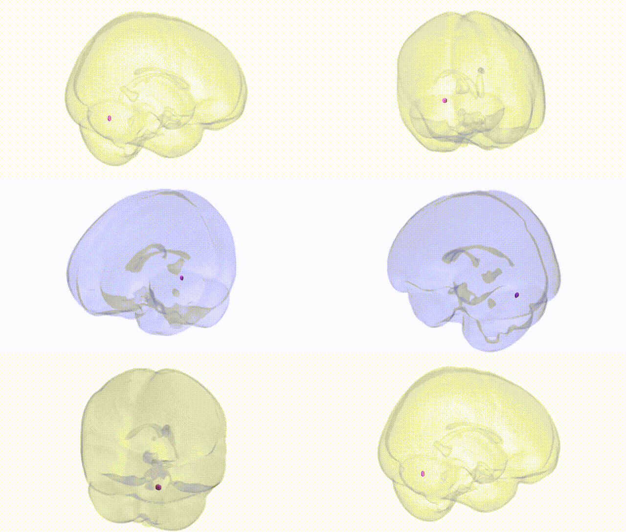
Full Quality Version: Download MP4
Wali Sidiqyar*, Gaurav Rudravaram*, Elyssa M. McMaster, Trent M. Schwartz, Adam M. Saunders, Kurt G. Schilling, Bennett A. Landman "Introducing SPINS: A Shared Public Visualization Library of Neuroanatomical Structures." Medical Imaging with Deep Learning- short paper
This resource is licensed under CC0 1.0 Universal (Public Domain).