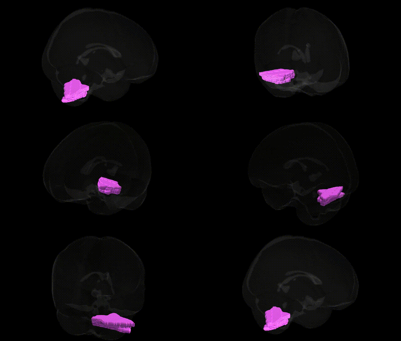
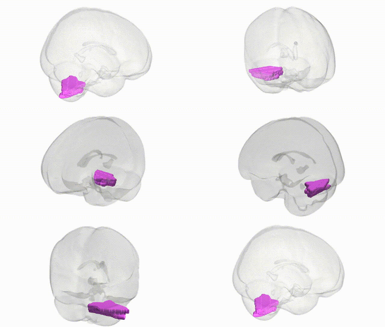
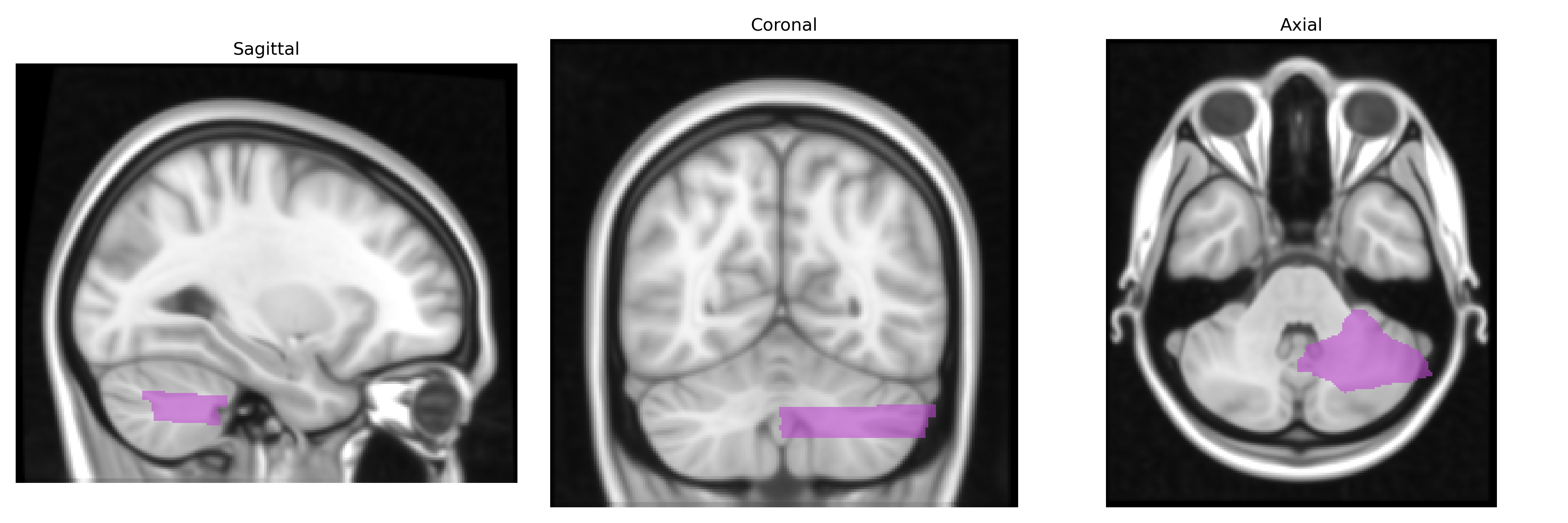
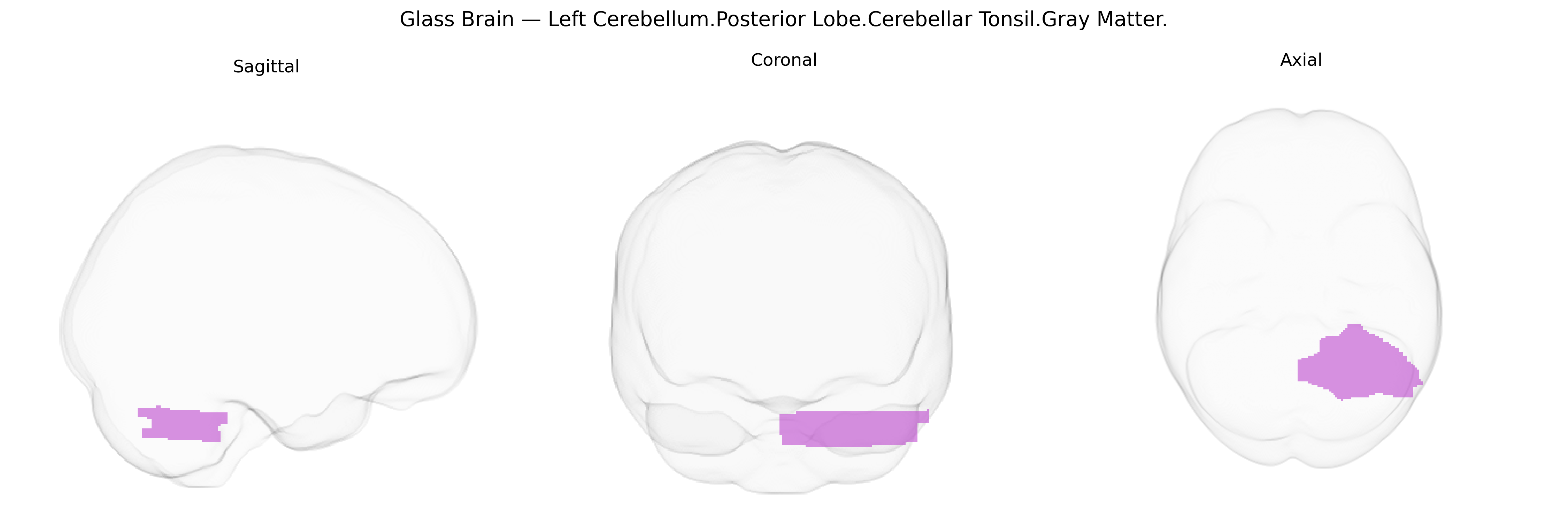
The bilateral Left Cerebellum.Posterior Lobe.Cerebellar Tonsil.Gray Matter corresponds to the gray matter of the cerebellar tonsils, paired ovoid lobules located on the inferior surface of the cerebellar hemispheres and forming part of the posterior lobe. These structures participate in the modulation of motor coordination, balance, and posture by integrating proprioceptive and vestibular inputs and refining descending motor commands via connections with deep cerebellar nuclei and brainstem pathways. The cerebellar tonsils are clinically important because downward displacement through the foramen magnum can occur in conditions such as Chiari malformations or posterior fossa mass lesions, leading to compression of the medulla and disruption of cardiorespiratory and motor functions. There is no direct link for this exact subdivision; a related structure is the Cerebellum.
The bilateral left posterior cerebellar tonsil gray matter (Talairach “Left Cerebellum.Posterior Lobe.Cerebellar Tonsil.Gray Matter”) is part of the posteroinferior cerebellum, a region implicated in coordination, affective regulation, and cognitive processes, and although relatively few imaging-genetics or GWAS studies isolate the tonsil specifically, converging evidence from cerebellar structural and functional genomics implicates several pathways and traits. Large-scale brain-imaging GWAS (e.g., ENIGMA, UK Biobank) have identified common variants in genes related to neurodevelopment, synaptic function, and axon guidance (including loci near genes such as KIAA0586, RELN, and DLG2) that influence overall cerebellar volume and posterior lobe morphology, with downstream effects on regional gray-matter thickness and surface area that encompass the tonsillar territory. Polygenic overlap has been reported between cerebellar volume and neuropsychiatric or neurodevelopmental conditions such as schizophrenia, major depressive disorder, autism spectrum disorder, and ADHD, as well as cognitive traits and educational attainment; in these studies, cerebellar posterior lobe and vermis/tonsillar regions frequently show gray-matter or connectivity alterations linked to genetic liability. Additionally, rare variant and copy-number analyses in disorders with cerebellar malformations (e.g., Dandy–Walker malformation, Joubert syndrome, some forms of spinocerebellar ataxia) implicate genes involved in ciliogenesis, transcriptional regulation, and ion channels that can affect posterior fossa development and cerebellar tonsil position or volume, while common variants associated with migraine, balance and vestibular traits, and essential tremor have been connected to cerebellar circuits that include the tonsils. Overall, the genetic architecture of this region appears highly polygenic and shared with broader cerebellar and cortico-cerebellar networks, with no single variant or gene exclusively defining the bilateral left posterior cerebellar tonsil gray matter but rather a distributed set of neurodevelopmental and synaptic genes contributing to its structure and function.
Overview generated by GPT-4o (2026).
Region ID: 3
Hemisphere: bilateral
Atlas: Talairach labels 1mm
Full Quality Version: Download MP4
Full Quality Version: Download MP4
Wali Sidiqyar*, Gaurav Rudravaram*, Elyssa M. McMaster, Trent M. Schwartz, Adam M. Saunders, Kurt G. Schilling, Bennett A. Landman "Introducing SPINS: A Shared Public Visualization Library of Neuroanatomical Structures." Medical Imaging with Deep Learning- short paper
This resource is licensed under CC0 1.0 Universal (Public Domain).