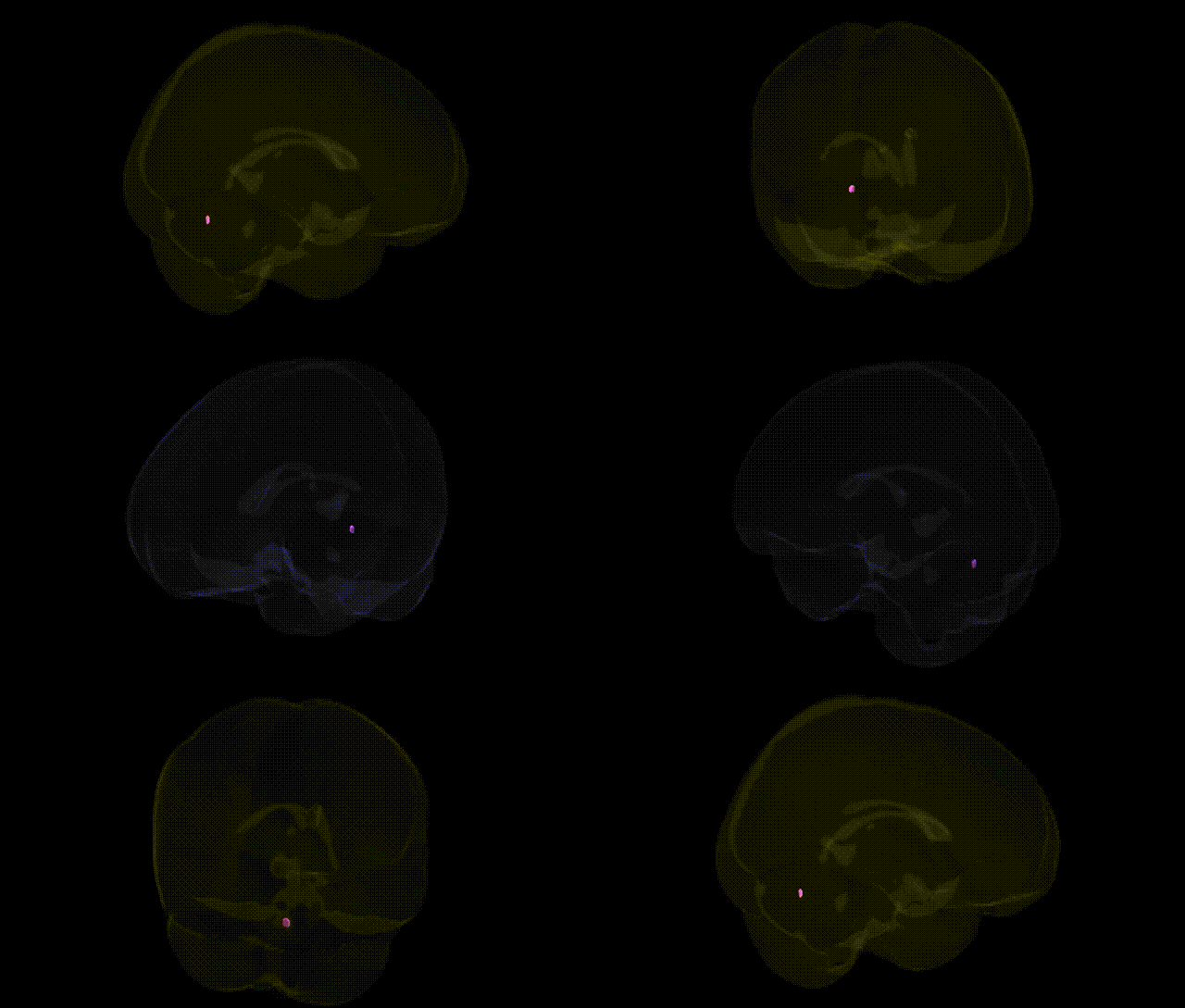
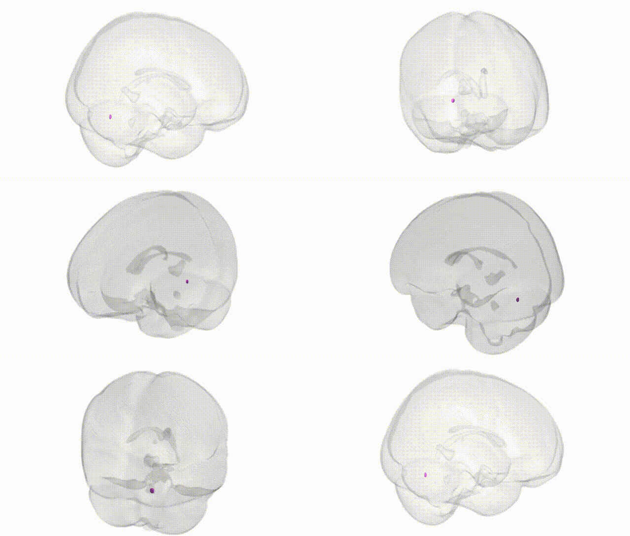
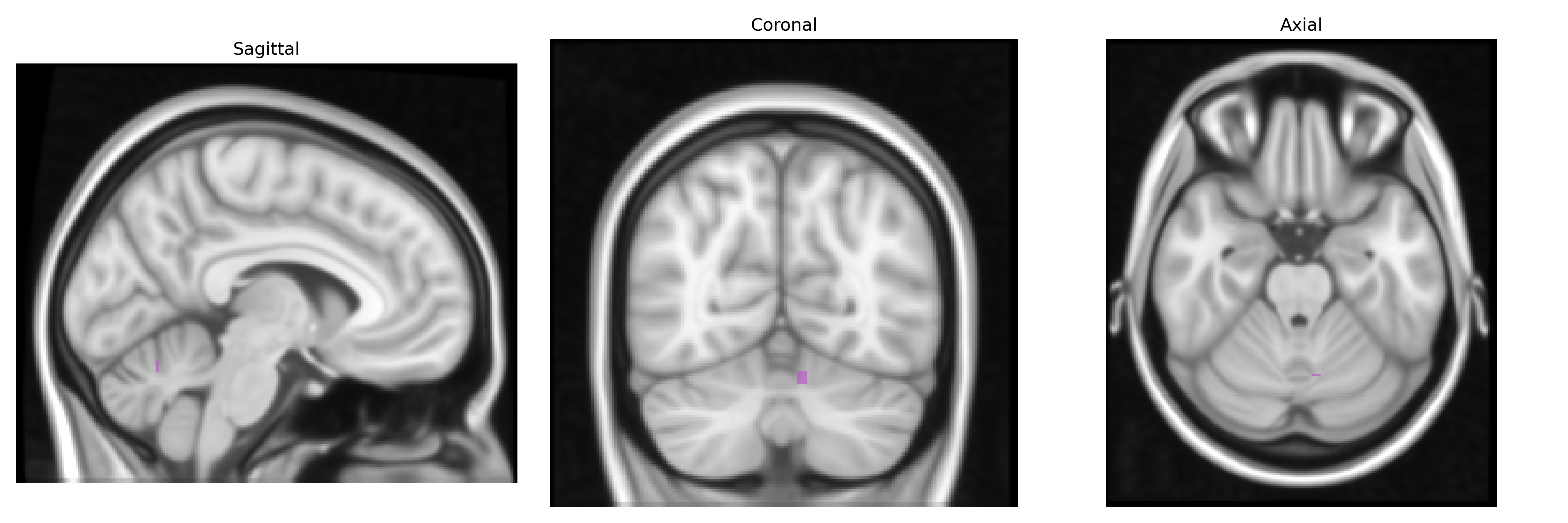
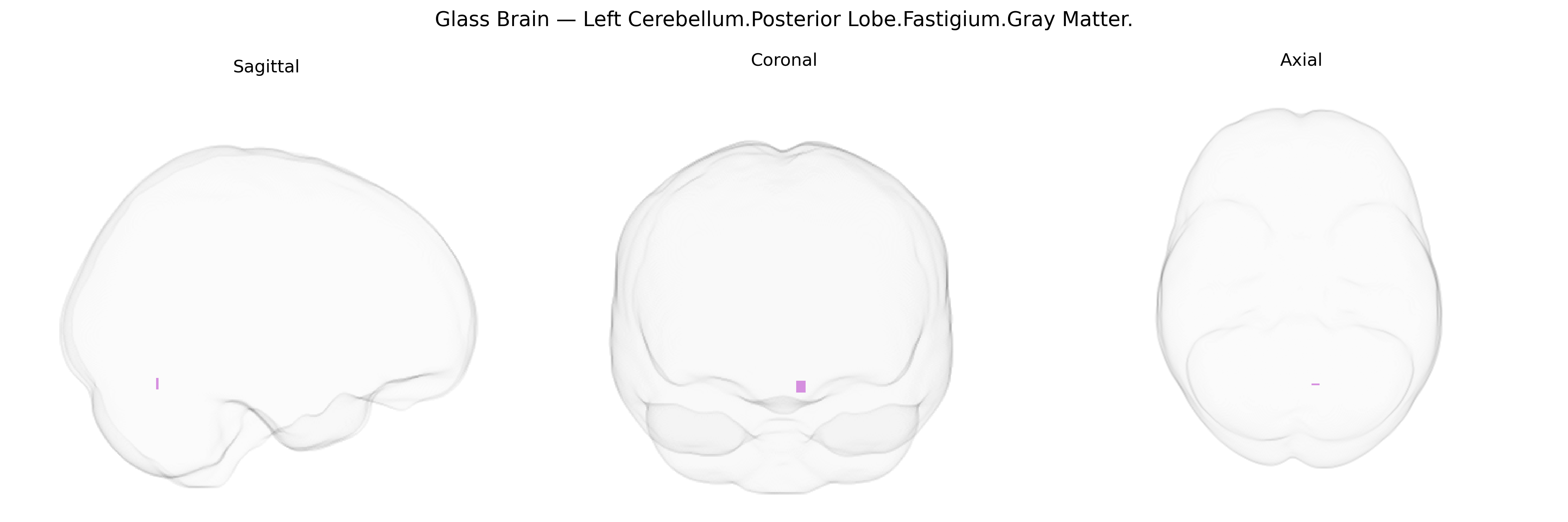
The bilateral Left Cerebellum Posterior Lobe Fastigium Gray Matter corresponds to gray matter within the fastigial region of the posterior lobe of the cerebellum, situated near the midline in the roof of the fourth ventricle and associated with the fastigial (medial cerebellar) nucleus. This region participates in integrating vestibular, proprioceptive, and somatosensory information to modulate axial and proximal limb musculature, contributing to posture, balance, and gait control. Functionally, it is part of the vestibulocerebellar and spinocerebellar networks, receiving input from spinal and vestibular pathways and projecting to vestibular nuclei and reticular formation, thereby influencing muscle tone and eye–head coordination. There is no direct link for this precise subregion; a related structure is the Cerebellum.
The bilateral left cerebellar posterior lobe fastigium gray matter (Talairach 1 mm atlas) lies within cerebellar midline/posterior territory that has been implicated in several genetic and GWAS-based associations, although few studies target the fastigium specifically; instead, it is typically subsumed within broader posterior cerebellar or vermal regions. Large imaging–genetics consortia (e.g., ENIGMA, UK Biobank) have shown that cerebellar gray matter volume and lobular morphology, including posterior lobe and vermis regions encompassing or adjacent to the fastigial area, exhibit significant SNP-based heritability and polygenic influences, with loci near genes involved in neurodevelopment, synaptic function, and axon guidance (such as variants in LINGO1, DCC, and ROBO family genes) contributing to interindividual differences in cerebellar structure. Mendelian and rare-variant studies of cerebellar malformations (e.g., mutations in genes like FOXC1, PTF1A, and various Joubert-related genes) can produce hypoplasia or dysplasia that includes posterior vermis/fastigial regions. Functionally, posterior cerebellar and vermal territories including or bordering the fastigium have been genetically and structurally linked to neurodevelopmental and psychiatric conditions such as autism spectrum disorder, schizophrenia, bipolar disorder, and major depression, as well as to motor coordination phenotypes and essential tremor, in which risk loci often map to genes regulating cerebellar circuitry and Purkinje cell function. Moreover, imaging–GWAS and polygenic score studies have connected interindividual variation in posterior cerebellar gray matter to cognitive traits (working memory, processing speed, general intelligence) and affective traits (neuroticism, anxiety-related measures), suggesting that the genetic architecture shaping the fastigial/posterior cerebellar region contributes to both motor and non-motor domains, even though precise fastigium-specific GWAS signals remain scarce due to current atlas resolutions and region-of-interest definitions.
Overview generated by GPT-4o (2026).
Region ID: 162
Hemisphere: bilateral
Atlas: Talairach labels 1mm
Full Quality Version: Download MP4
Full Quality Version: Download MP4
Wali Sidiqyar*, Gaurav Rudravaram*, Elyssa M. McMaster, Trent M. Schwartz, Adam M. Saunders, Kurt G. Schilling, Bennett A. Landman "Introducing SPINS: A Shared Public Visualization Library of Neuroanatomical Structures." Medical Imaging with Deep Learning- short paper
This resource is licensed under CC0 1.0 Universal (Public Domain).