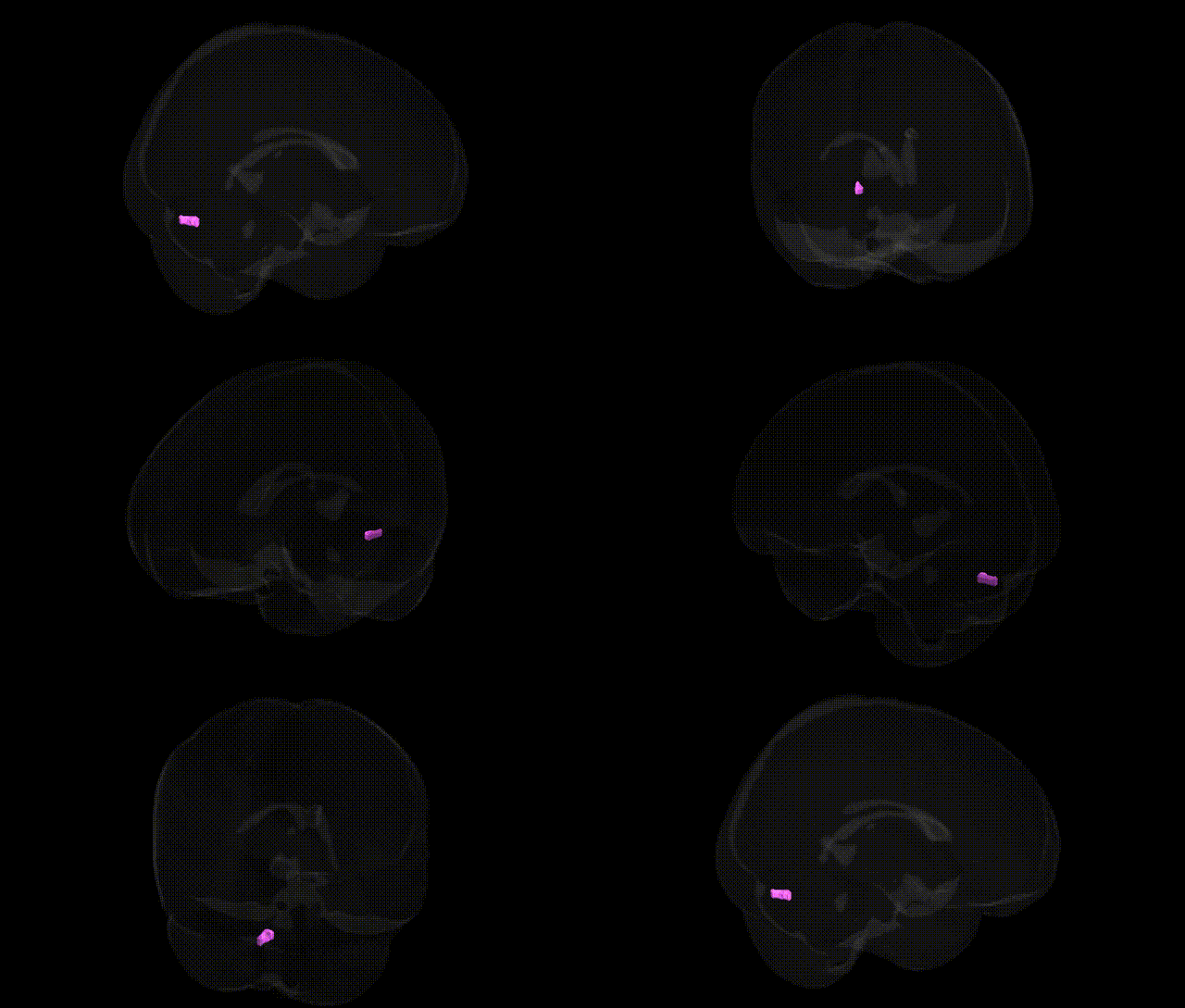
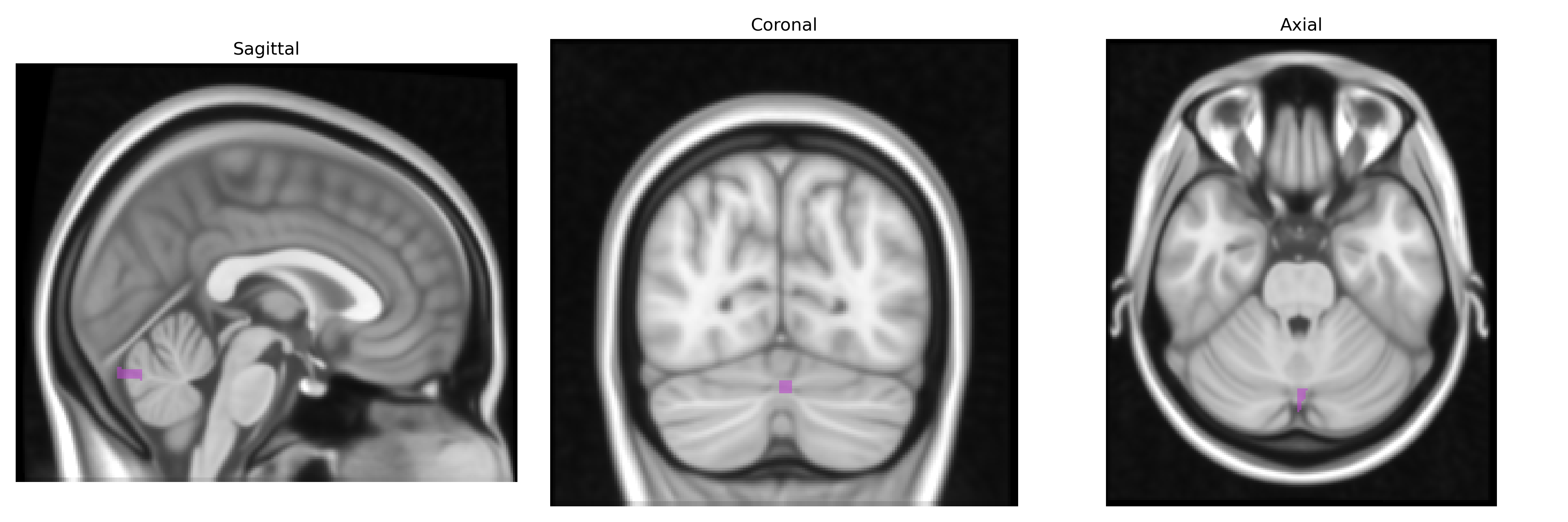
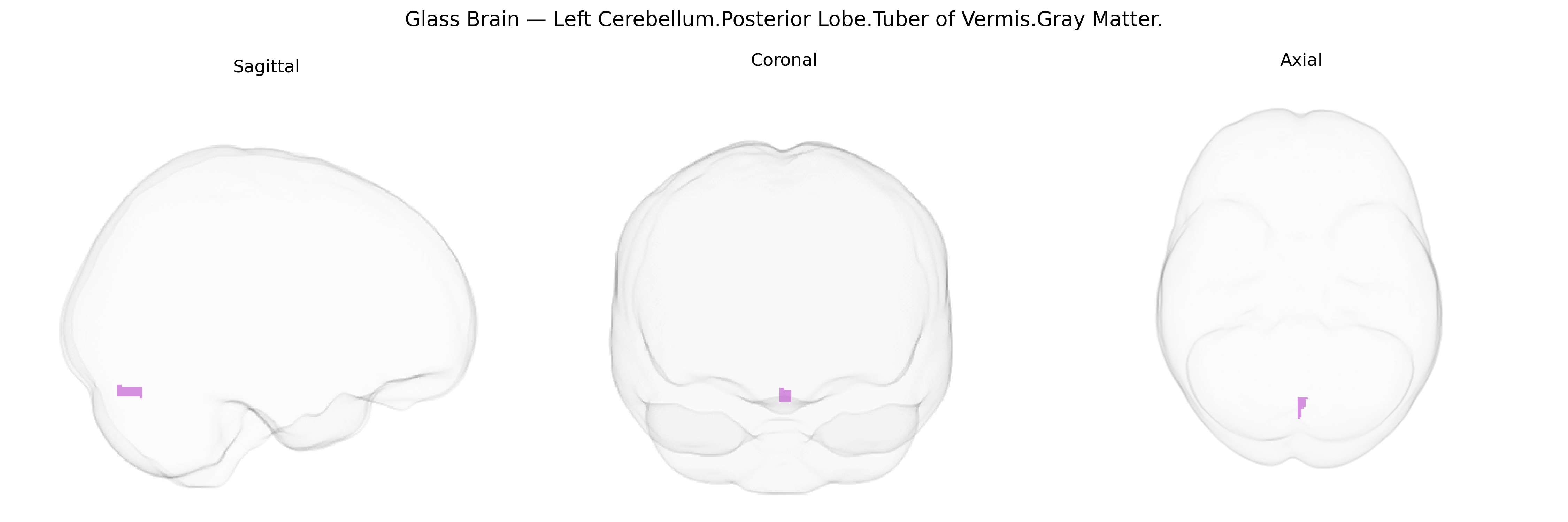
The bilateral Left Cerebellum Posterior Lobe, Tuber of Vermis, Gray Matter corresponds to part of lobule VII of the cerebellar vermis, a midline structure of the posterior lobe involved in the coordination of trunk and proximal limb movements, as well as aspects of posture and balance. The tuber of the vermis lies between the hemispheric cerebellar lobules and contributes to integrating somatosensory and motor information to fine-tune ongoing movements. Functionally, the posterior lobe, including this region, is also implicated in higher-order processes such as motor learning and, to some extent, cognitive and affective modulation through its connections with cortical and subcortical structures. There is no direct link for this specific subregion; a related article is: Cerebellar vermis.
The bilateral Left Cerebellum.Posterior Lobe.Tuber of Vermis.Gray Matter region, as defined in the Talairach labels 1 mm atlas, lies within the posterior vermis, a cerebellar zone repeatedly implicated in genetic studies of neurodevelopmental and psychiatric conditions rather than in region-specific GWAS hits per se. Large brain-imaging GWAS (e.g., ENIGMA, UK Biobank) have shown that gray matter volume and cortical thickness in posterior cerebellar regions, including vermis and adjacent lobules, are influenced by common variants in genes involved in neurodevelopment (e.g., MAPT region on 17q21, FOXP1/FOXP2-related pathways, and polygenic architectures overlapping with general brain volume loci), though these are typically reported at the level of “cerebellar gray matter” or specific lobules rather than the tuber of the vermis alone. Genetic association and linkage studies in autism spectrum disorder, schizophrenia, and bipolar disorder have linked risk variants in synaptic and neurodevelopmental genes (such as RELN, CNTNAP2, SHANK family genes) to structural and functional abnormalities in the cerebellar vermis, with reduced or altered vermal volumes frequently reported in carriers of higher polygenic risk for these disorders. Additionally, cerebellar vermis morphology has been associated with genetic liability to attention-deficit/hyperactivity disorder, mood disorders, and some forms of ataxia and developmental coordination disorders, where pathogenic or likely pathogenic variants in ion channel, mitochondrial, and cerebellar patterning genes (e.g., CACNA1A, ATXN genes, and others) show convergent effects on midline cerebellar structures. Overall, current evidence supports a polygenic and pleiotropic architecture in which variants influencing neurodevelopment, synaptic function, and brain-wide morphometry contribute to interindividual differences in posterior vermis gray matter, but no single variant or locus has been uniquely or consistently assigned specifically to the Left Cerebellum.Posterior Lobe.Tuber of Vermis.Gray Matter label.
Overview generated by GPT-4o (2026).
Region ID: 114
Hemisphere: bilateral
Atlas: Talairach labels 1mm
Full Quality Version: Download MP4
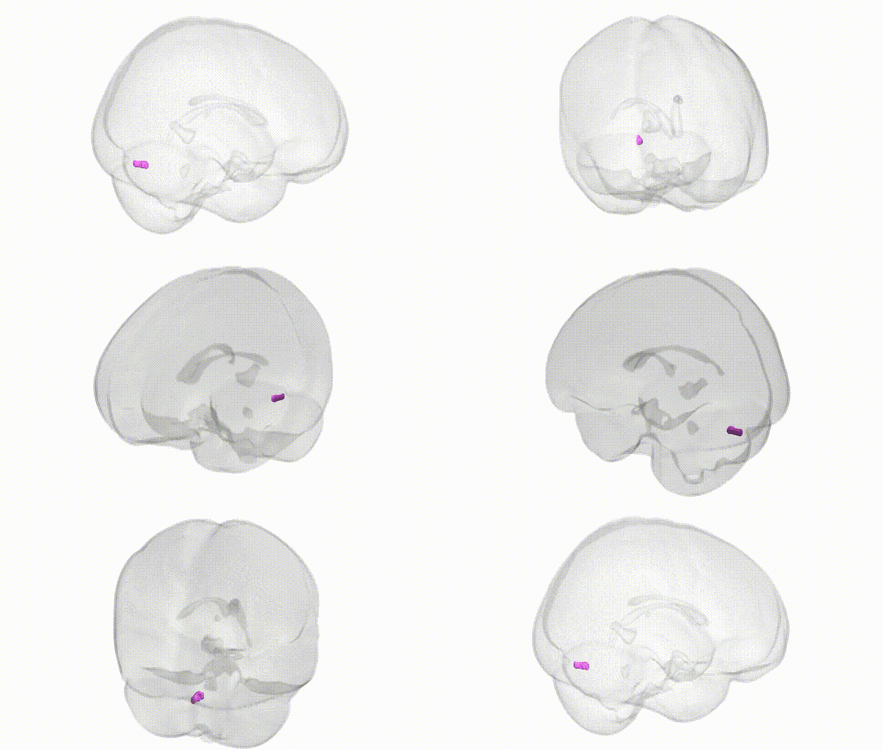
Full Quality Version: Download MP4
Wali Sidiqyar*, Gaurav Rudravaram*, Elyssa M. McMaster, Trent M. Schwartz, Adam M. Saunders, Kurt G. Schilling, Bennett A. Landman "Introducing SPINS: A Shared Public Visualization Library of Neuroanatomical Structures." Medical Imaging with Deep Learning- short paper
This resource is licensed under CC0 1.0 Universal (Public Domain).