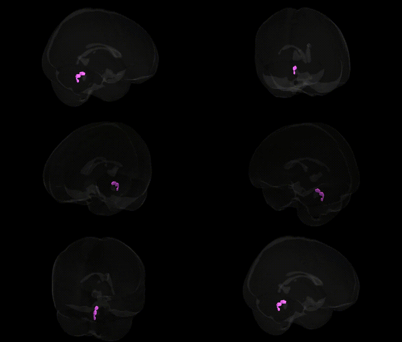
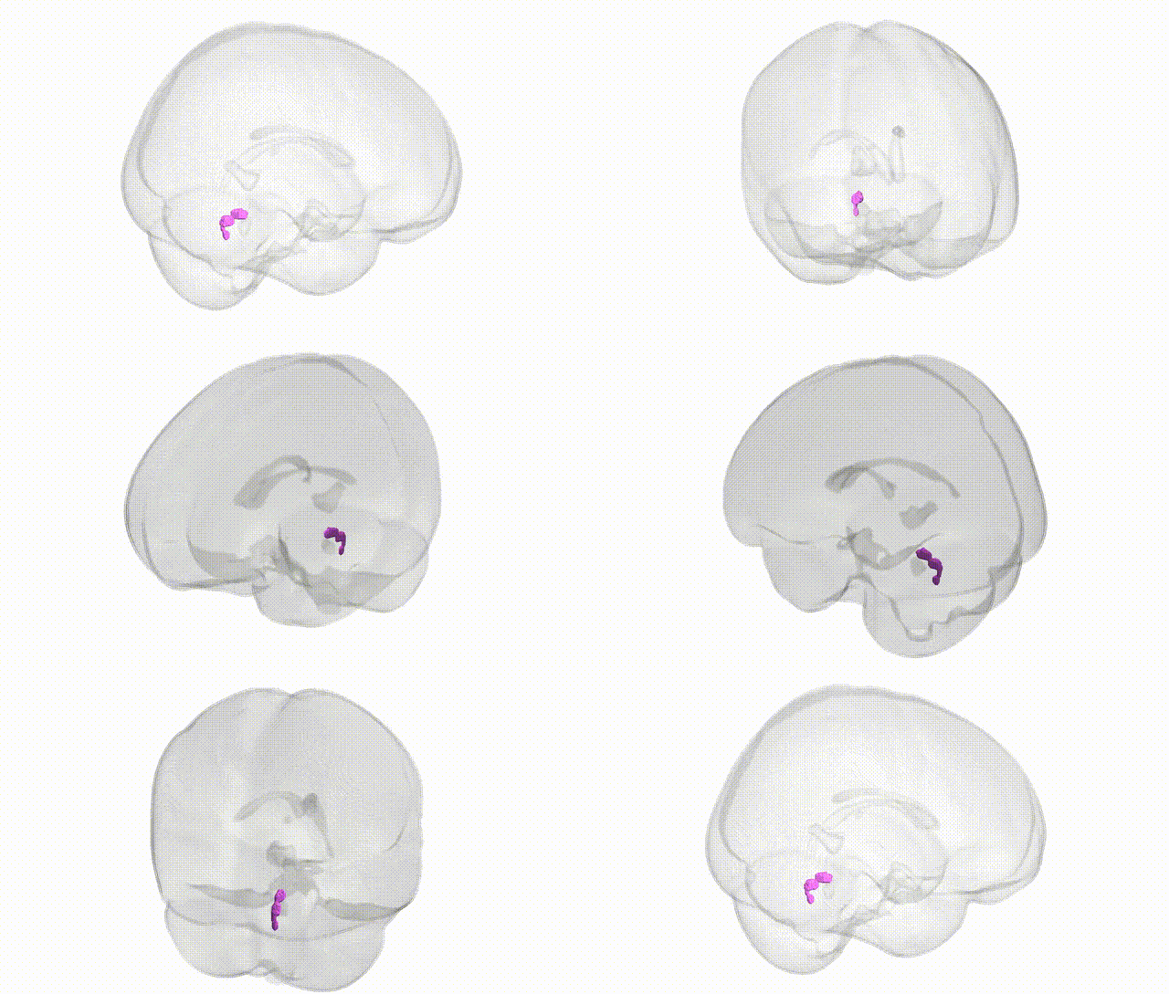
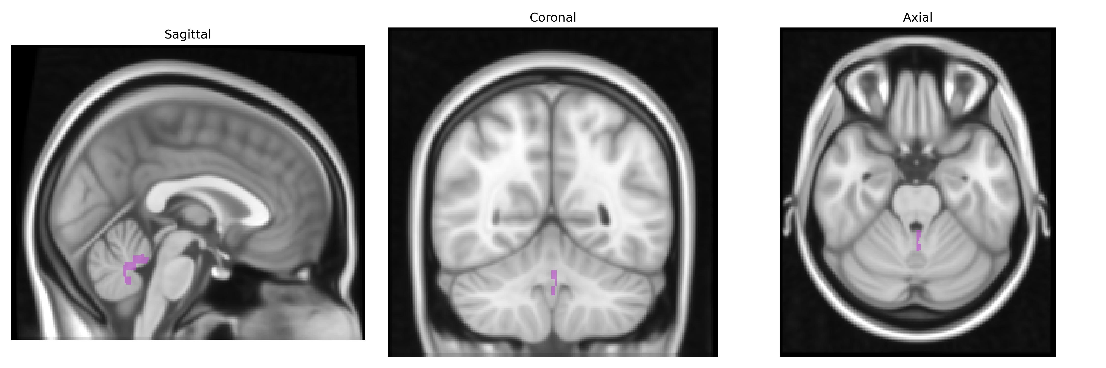
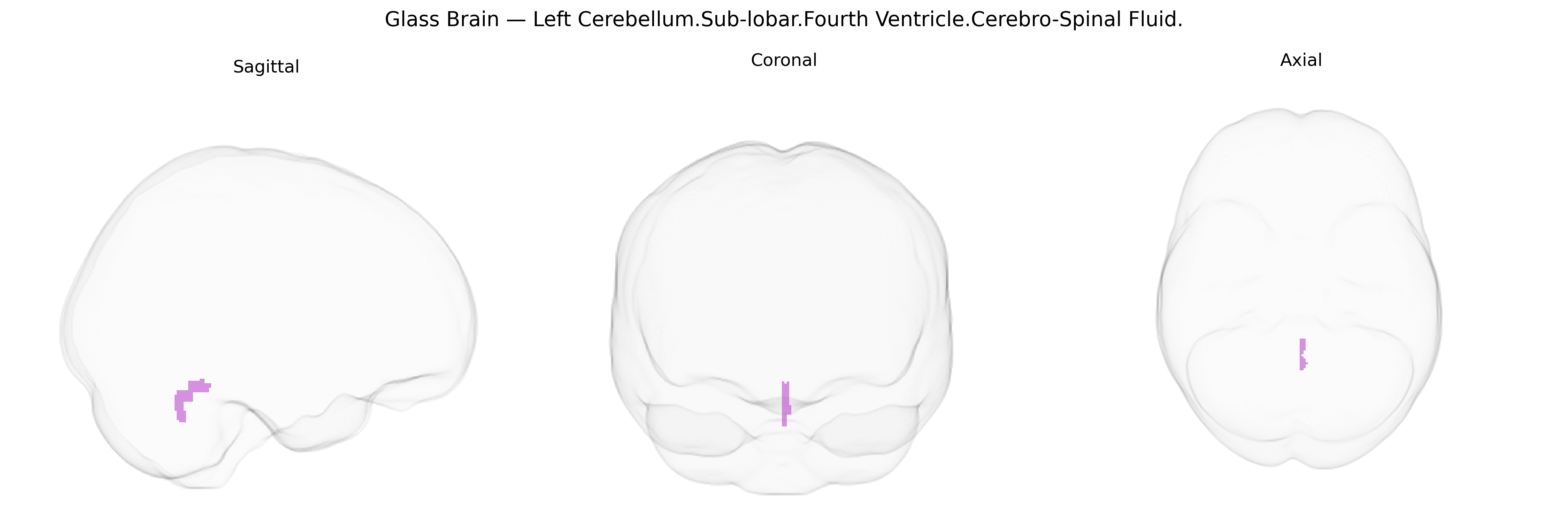
The bilateral Left Cerebellum.Sub-lobar.Fourth Ventricle.Cerebro-Spinal Fluid region, as labeled in the Talairach 1 mm atlas, corresponds primarily to the cerebrospinal fluid (CSF)–filled lumen of the fourth ventricle, which lies dorsal to the pons and medulla and ventral to the cerebellum, extending across the midline and into left and right subregions for atlas labeling purposes. This space forms part of the ventricular system that circulates CSF, contributing to mechanical cushioning of the brain, maintenance of intracranial pressure, and regulation of the chemical environment surrounding neural tissue. Inferiorly, the fourth ventricle continues into the central canal of the spinal cord and communicates with the subarachnoid space via the median and lateral apertures, while superiorly it narrows into the cerebral aqueduct. In the Talairach framework, voxels labeled in this region reflect fluid rather than gray or white matter, and functionally they are interpreted in relation to CSF dynamics and surrounding brainstem and cerebellar structures rather than as a discrete neural nucleus or cortex.
Genetic association findings specific to the bilateral “Left Cerebellum.Sub-lobar.Fourth Ventricle.Cerebro-Spinal Fluid” label from the Talairach 1 mm atlas are sparse, as most imaging genetics and GWAS studies treat the cerebellar cortex, deep nuclei, and cerebrospinal fluid (CSF) spaces more broadly rather than this precise parcellation. Large brain-structure GWAS (e.g., ENIGMA and UK Biobank–based studies) have identified associations between cerebellar volume and variants in genes related to neurodevelopment, axon guidance, and synaptic function (such as loci near KIAA0586/TALPID3, PAX3, and several 3p21 and 6q regions), while ventricular and total CSF volumes show robust links to risk alleles for Alzheimer’s disease and related neurodegeneration (including APOE, BIN1, CLU, CR1), as well as schizophrenia and bipolar disorder risk loci that influence global brain and ventricular size. CSF volume and fourth ventricle size are also indirectly associated, through polygenic scores, with traits such as general cognitive ability, intracranial volume, and height, reflecting shared developmental and growth pathways. However, no major GWAS has isolated a distinct genetic signal uniquely attributable to the Talairach-defined fourth ventricle CSF region, and reported associations are best understood as reflecting genetic influences on overall cerebellar morphology, posterior fossa development, and global CSF/ventricular expansion rather than on this specific atlas parcel.
Overview generated by GPT-4o (2026).
Region ID: 70
Hemisphere: bilateral
Atlas: Talairach labels 1mm
Full Quality Version: Download MP4
Full Quality Version: Download MP4
Wali Sidiqyar*, Gaurav Rudravaram*, Elyssa M. McMaster, Trent M. Schwartz, Adam M. Saunders, Kurt G. Schilling, Bennett A. Landman "Introducing SPINS: A Shared Public Visualization Library of Neuroanatomical Structures." Medical Imaging with Deep Learning- short paper
This resource is licensed under CC0 1.0 Universal (Public Domain).