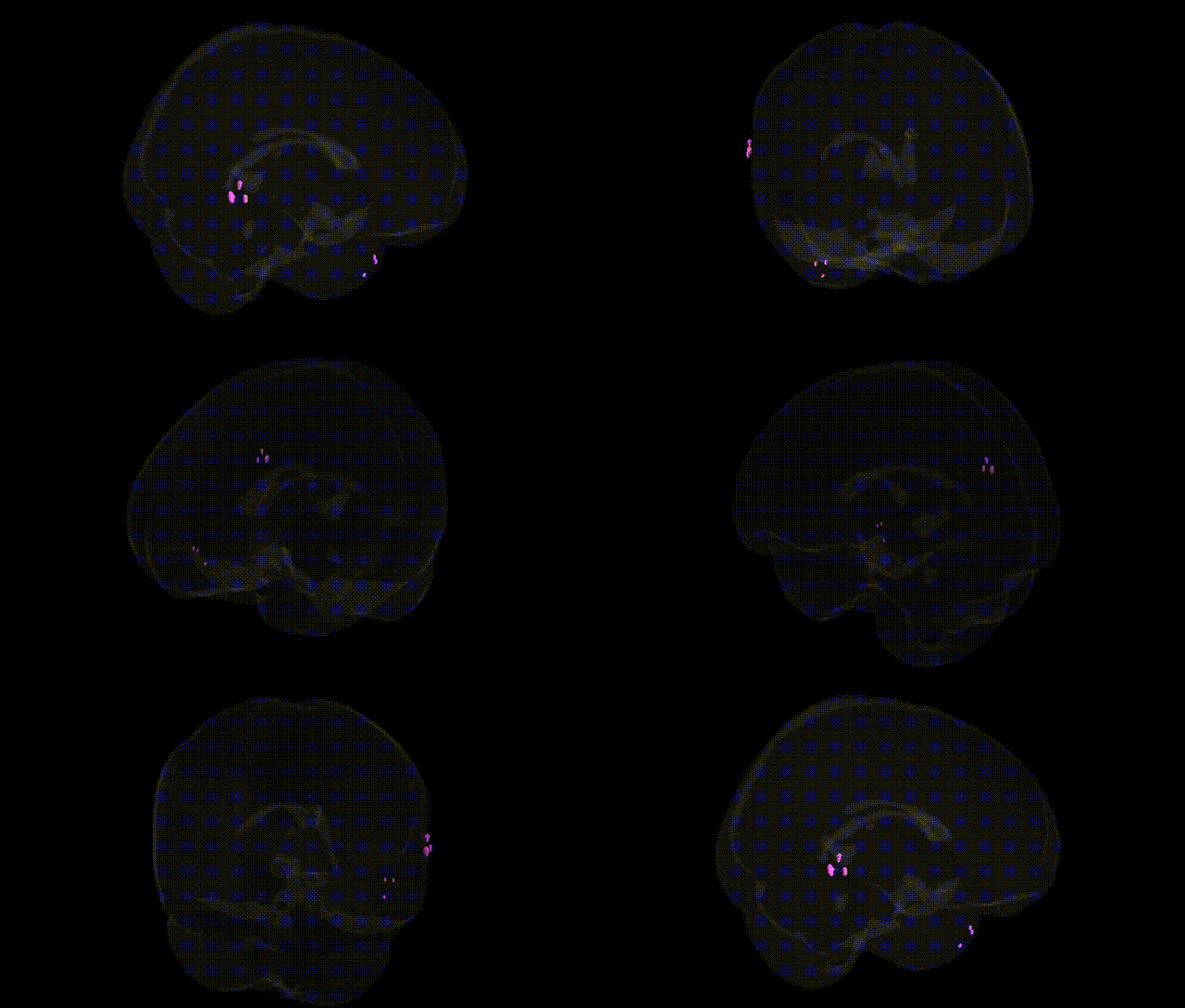
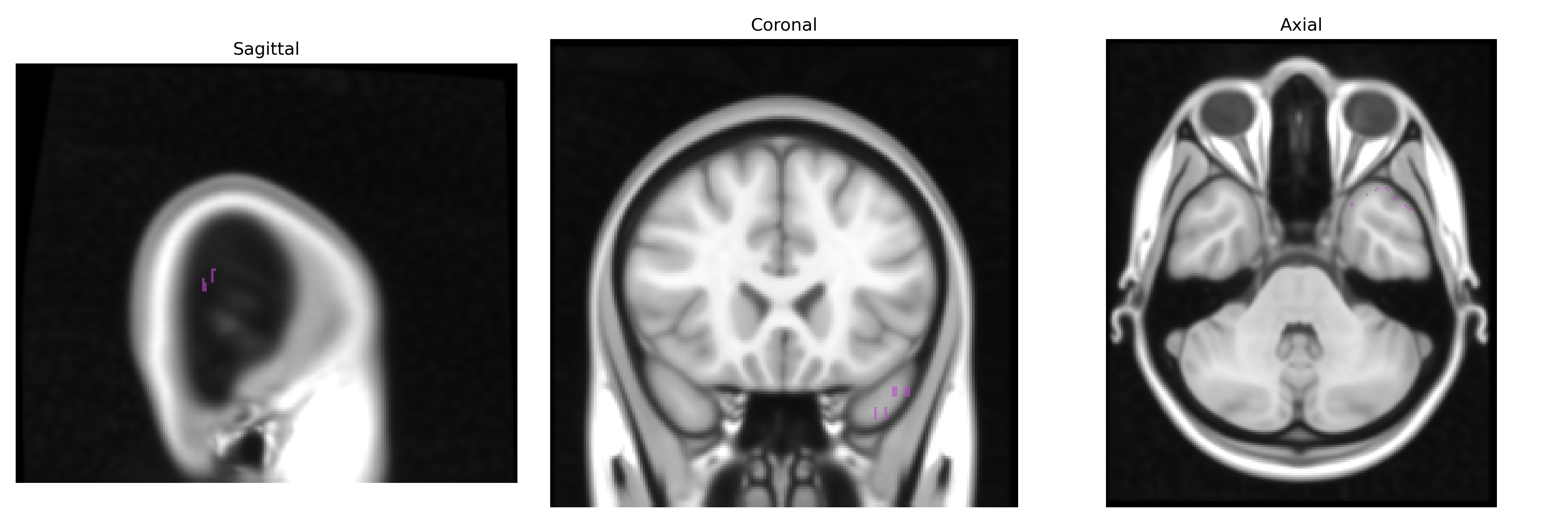
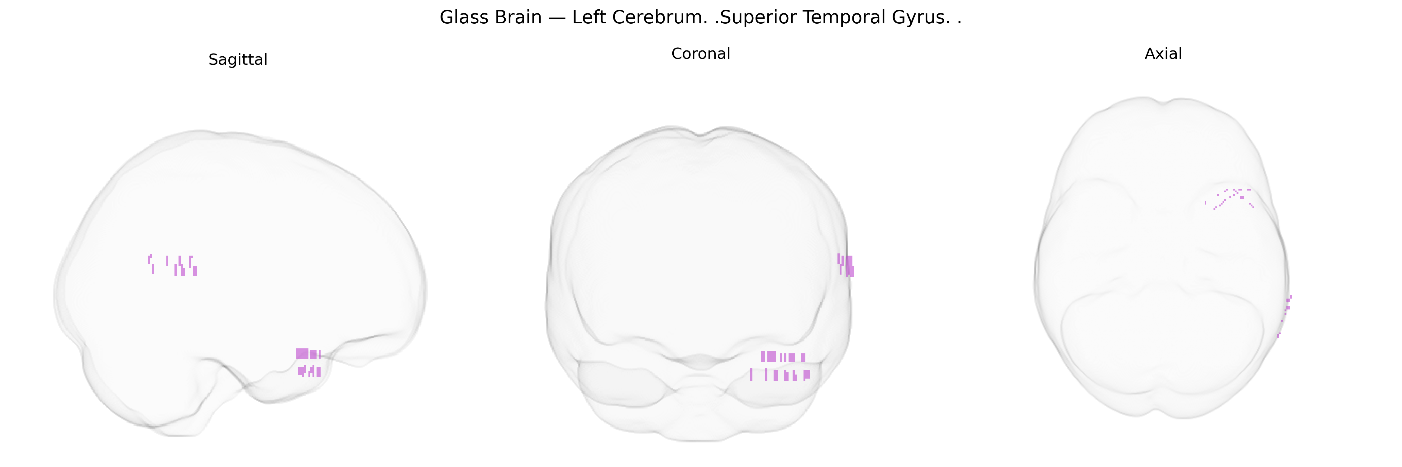
The bilateral Left Cerebrum Superior Temporal Gyrus, as defined in the Talairach 1 mm Atlas, corresponds to cortical tissue along the superior aspect of the temporal lobe, extending from the temporal pole posteriorly toward the parietal lobe and bordering the lateral sulcus (Sylvian fissure) superiorly. This region encompasses primary and secondary auditory cortices, including Heschl’s gyrus on the superior temporal plane, and is critically involved in processing complex acoustic information, speech perception, language comprehension (notably in the posterior portion overlapping Wernicke’s area in the dominant hemisphere), and aspects of social cognition such as voice recognition and interpretation of prosody. Cytoarchitectonically, it includes Brodmann areas predominantly within BA 22 and adjacent regions, receiving multimodal input from auditory, parietal, and frontal association areas and projecting to widespread cortical and subcortical targets that support auditory-linguistic integration and higher-order temporal lobe functions. Superior temporal gyrus
The bilateral left superior temporal gyrus (STG) has been repeatedly implicated in imaging–genetics and GWAS studies of brain structure and function, particularly for cortical thickness and surface area, language and auditory processing, and psychiatric and neurodevelopmental disorders. Large-scale consortia such as ENIGMA and UK Biobank have identified common variants in genes involved in neurodevelopment (e.g., microtubule, axon guidance, and synaptic pathways) associated with STG morphology, including loci near or within genes such as MIR124-1, LAMC1, and others contributing to global and regional cortical measures, although specific Talairach-based STG foci are rarely isolated. Variants in FOXP2 and related language network genes have been linked to structural and functional differences in the superior temporal cortex, consistent with its role in speech perception and comprehension, while polygenic risk for schizophrenia, bipolar disorder, and major depression is associated with reduced STG volume or altered cortical thickness, reflecting convergent genetic influences on auditory hallucinations, language disturbances, and affective dysregulation. Autism spectrum disorder and specific language impairment GWAS and candidate-gene studies also implicate genes affecting synaptic plasticity and cortical patterning (e.g., CNTNAP2, NRXN1, and other neurodevelopmental loci) that modulate STG structure and connectivity, and auditory cortex–focused analyses highlight associations between genetic variants shaping excitatory–inhibitory balance and altered superior temporal function in disorders such as dyslexia and epilepsy, although effect sizes remain modest and highly polygenic.
Overview generated by GPT-4o (2026).
Region ID: 100
Hemisphere: bilateral
Atlas: Talairach labels 1mm
Full Quality Version: Download MP4
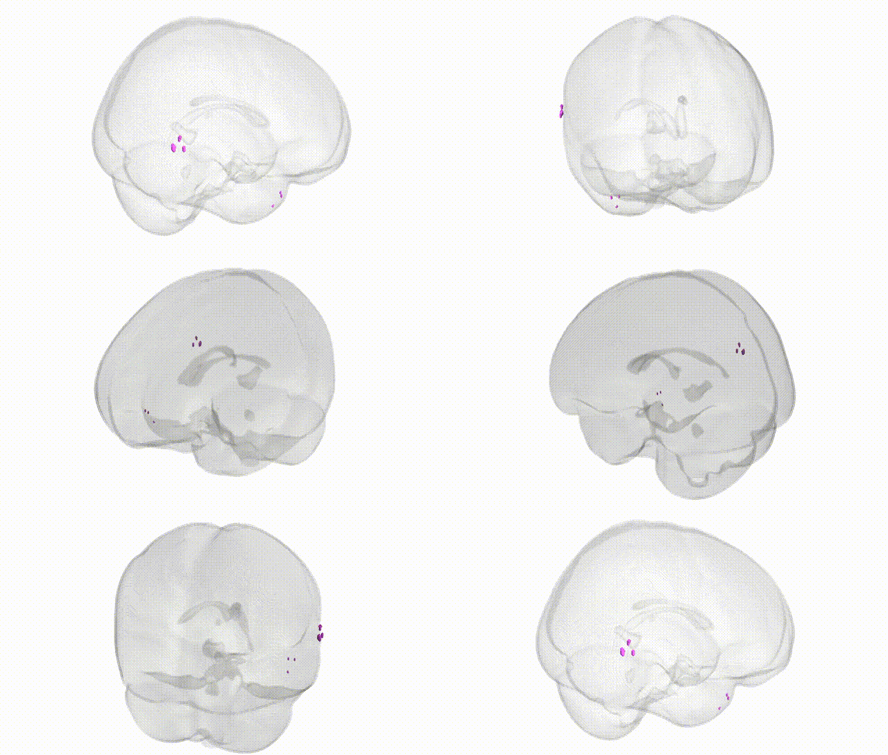
Full Quality Version: Download MP4
Wali Sidiqyar*, Gaurav Rudravaram*, Elyssa M. McMaster, Trent M. Schwartz, Adam M. Saunders, Kurt G. Schilling, Bennett A. Landman "Introducing SPINS: A Shared Public Visualization Library of Neuroanatomical Structures." Medical Imaging with Deep Learning- short paper
This resource is licensed under CC0 1.0 Universal (Public Domain).