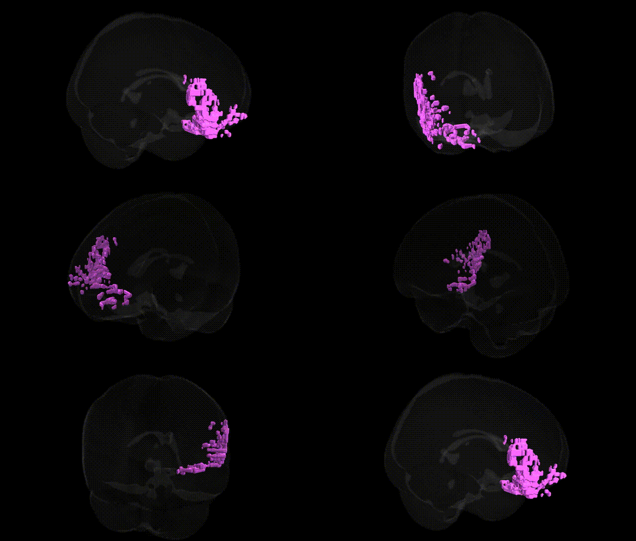
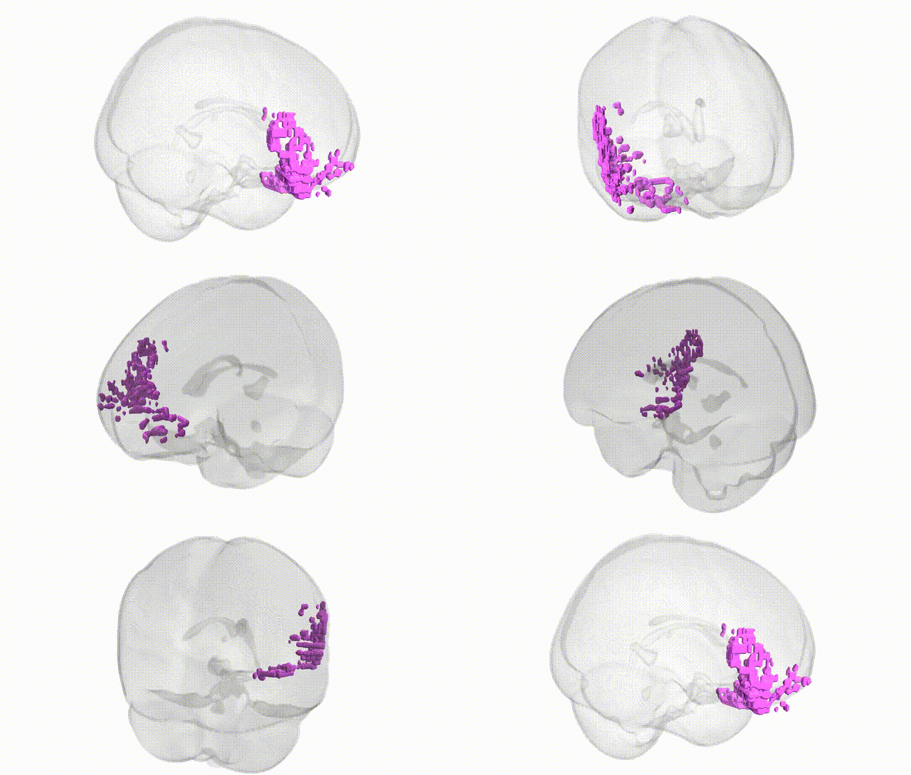
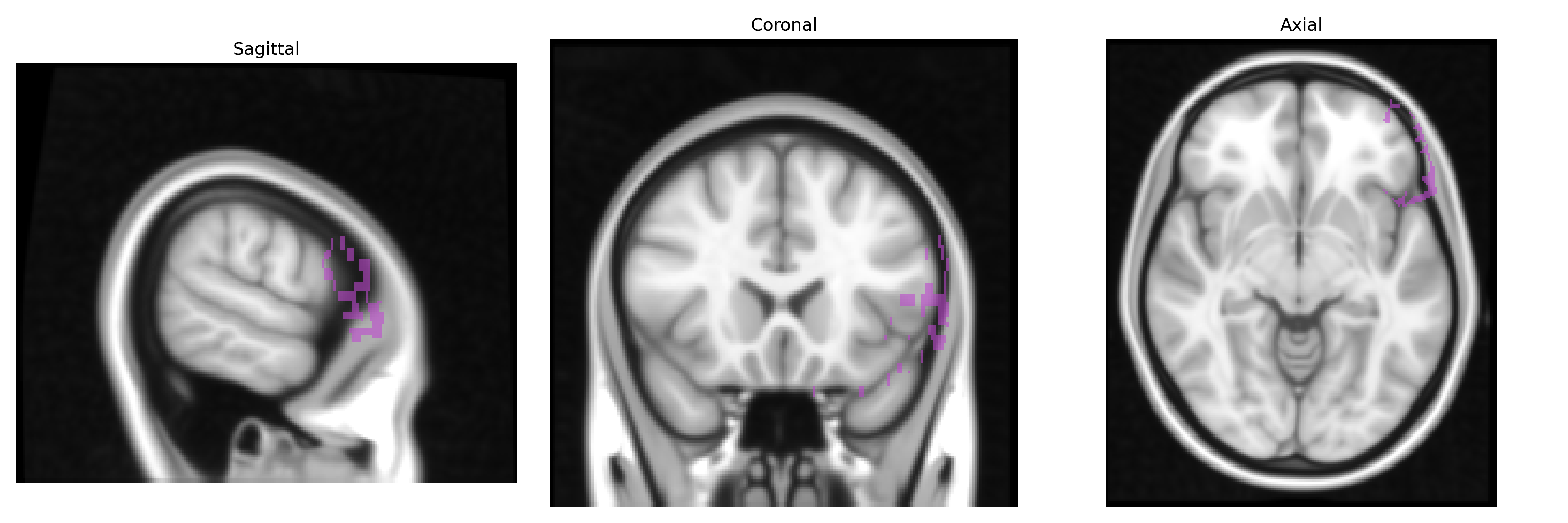
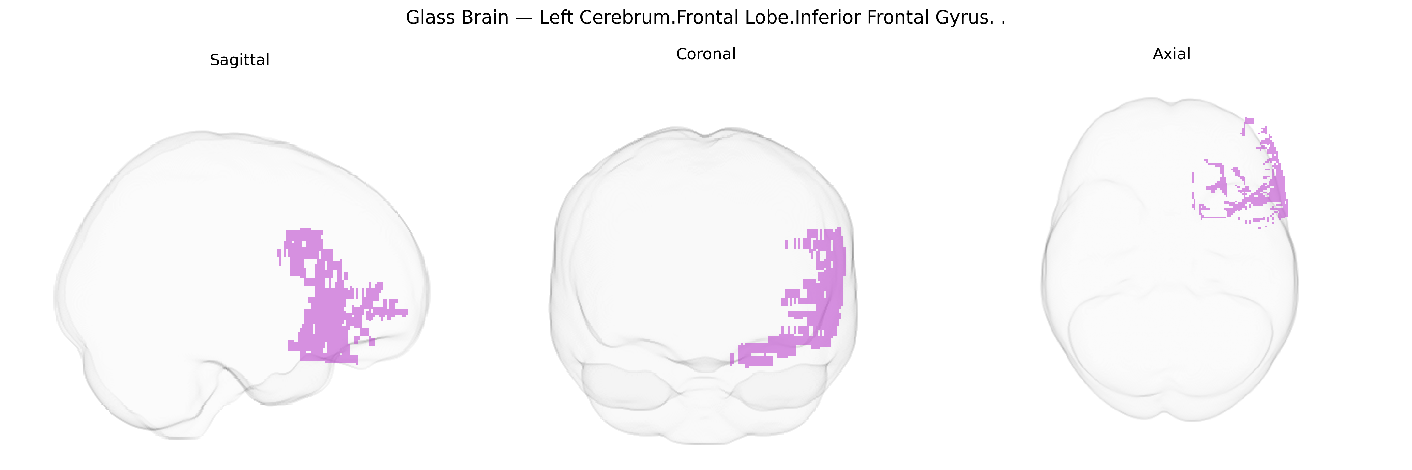
The bilateral Left Cerebrum.Frontal Lobe.Inferior Frontal Gyrus corresponds to a cortical region in the inferior portion of the frontal lobe, bordering the lateral fissure and typically subdivided into pars opercularis, pars triangularis, and pars orbitalis. In the left hemisphere it is classically associated with Broca’s area (particularly pars opercularis and pars triangularis), supporting speech production, syntactic processing, and aspects of language planning, while bilaterally it contributes to executive control, response inhibition, working memory, and aspects of social cognition. This region is heavily interconnected with other frontal, temporal, and parietal association areas, as well as subcortical structures, forming part of fronto-striatal and fronto-temporal networks important for goal-directed behavior and cognitive flexibility. Functionally and cytoarchitectonically, it corresponds most closely to what is generally termed the inferior frontal gyrus: Inferior frontal gyrus.
The bilateral inferior frontal gyrus (IFG) of the left cerebrum, as defined in Talairach 1 mm atlases, shows convergent genetic associations from imaging–genetics, GWAS, and disorder-related studies implicating frontal cortical development, synaptic plasticity, and language- and control-related circuitry. Twin and SNP-heritability studies indicate moderate to high heritability for IFG volume, cortical thickness, and functional activation during tasks of response inhibition, working memory, and language, with common variant polygenic influences overlapping those for global cortical surface area (e.g., MAPT, WNT signaling, and chromatin-remodeling loci reported in ENIGMA and UK Biobank–based cortical GWAS). The left IFG (including pars opercularis and pars triangularis/Broca’s area) is repeatedly implicated in genetic studies of speech and language: FOXP2 and CNTNAP2 variants have been associated with altered IFG structure or activation in developmental language disorder, autism, and language-related endophenotypes, while GWAS for educational attainment, reading ability, and verbal IQ show polygenic effects that partly localize to frontal language networks. Psychiatric and neurodevelopmental GWAS also implicate this region indirectly: polygenic risk for schizophrenia, bipolar disorder, major depression, ADHD, and autism has been associated with altered inferior frontal morphology and function, consistent with large-scale ENIGMA findings linking risk loci in genes such as CACNA1C, ZNF804A, and GRIN2A to prefrontal structural variation. In neurodegenerative and motor-speech conditions, genetic variants in C9orf72, GRN, and MAPT associated with frontotemporal dementia and primary progressive aphasia show preferential atrophy and functional disruption in left IFG, while GWAS for stuttering and apraxia of speech highlight networks that prominently include the inferior frontal cortex. Overall, genetic associations for the left IFG converge on polygenic influences that modulate frontal cortical architecture and connectivity, contributing to risk or resilience for language, executive control, psychiatric illness, and frontotemporal neurodegeneration.
Overview generated by GPT-4o (2026).
Region ID: 186
Hemisphere: bilateral
Atlas: Talairach labels 1mm
Full Quality Version: Download MP4
Full Quality Version: Download MP4
Wali Sidiqyar*, Gaurav Rudravaram*, Elyssa M. McMaster, Trent M. Schwartz, Adam M. Saunders, Kurt G. Schilling, Bennett A. Landman "Introducing SPINS: A Shared Public Visualization Library of Neuroanatomical Structures." Medical Imaging with Deep Learning- short paper
This resource is licensed under CC0 1.0 Universal (Public Domain).