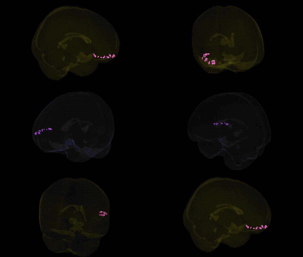
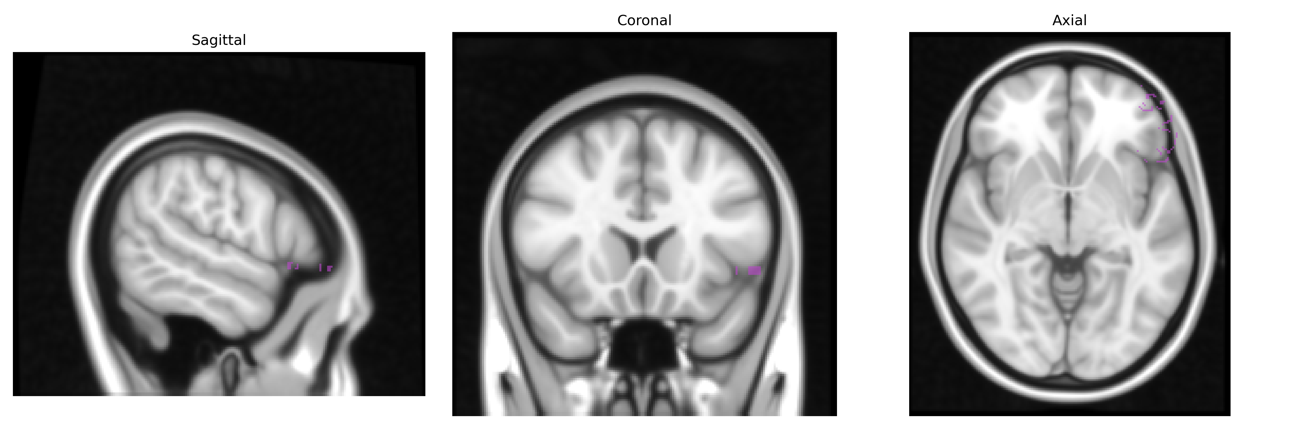
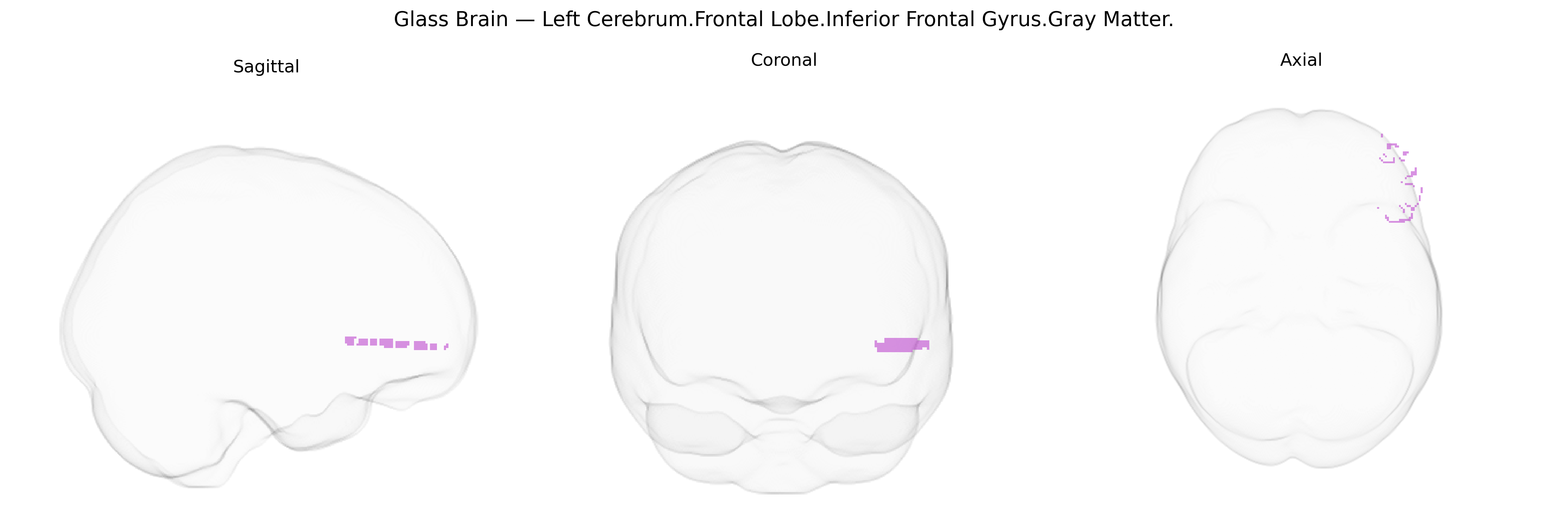
The bilateral left cerebrum frontal lobe inferior frontal gyrus gray matter corresponds primarily to cortical tissue in the pars opercularis, pars triangularis, and, to a lesser extent, pars orbitalis of the inferior frontal gyrus, encompassing regions classically associated with Broca’s area (typically Brodmann areas 44 and 45) in the dominant hemisphere. This region is heavily involved in speech and language production, phonological and syntactic processing, and aspects of semantic retrieval, as well as higher-order executive functions such as response inhibition, working memory, and cognitive control of behavior. It is richly interconnected with temporal and parietal association cortices, premotor and motor areas, and subcortical structures, forming part of the dorsal and ventral language streams and fronto-striatal networks. The underlying gray matter includes pyramidal neurons and interneurons arranged in distinct cortical layers, supporting complex integrative processing of linguistic, motor-planning, and cognitive signals. Inferior frontal gyrus
The bilateral left inferior frontal gyrus (IFG) gray matter, as defined in the Talairach 1 mm atlas, has been repeatedly implicated in genetic and genomic studies of language, cognitive control, and psychiatric risk, although specific GWAS hits are typically reported at the level of broader frontal or perisylvian regions rather than this exact parcel. Twin and family studies show high heritability for IFG volume and cortical thickness, with several large-scale imaging-genetics consortia (e.g., ENIGMA) identifying common variants in genes related to neuronal development, axon guidance, and synaptic plasticity (such as CNTNAP2, FOXP2-related pathways, and DISC1-related networks) that modulate inferior frontal structure and activation during language or working-memory tasks. Polygenic risk scores for schizophrenia, major depressive disorder, bipolar disorder, and attention-deficit/hyperactivity disorder have been associated with altered IFG gray matter volume or functional activation, and GWAS of reading, dyslexia, stuttering, and other speech/language traits similarly implicate left IFG as a key intermediate phenotype. In Alzheimer’s disease, frontotemporal dementia, and broader neurodegeneration, risk alleles (e.g., APOE ε4 and MAPT haplotypes) and GWAS-identified loci show associations with inferior frontal atrophy patterns, while autism spectrum condition genetics (including variants in synaptic and chromatin-remodeling genes) are linked to atypical IFG development and connectivity, supporting the role of this region as a convergence point where diverse genetic risks for language, executive dysfunction, and psychiatric disorders exert structural and functional effects.
Overview generated by GPT-4o (2026).
Region ID: 599
Hemisphere: bilateral
Atlas: Talairach labels 1mm
Full Quality Version: Download MP4
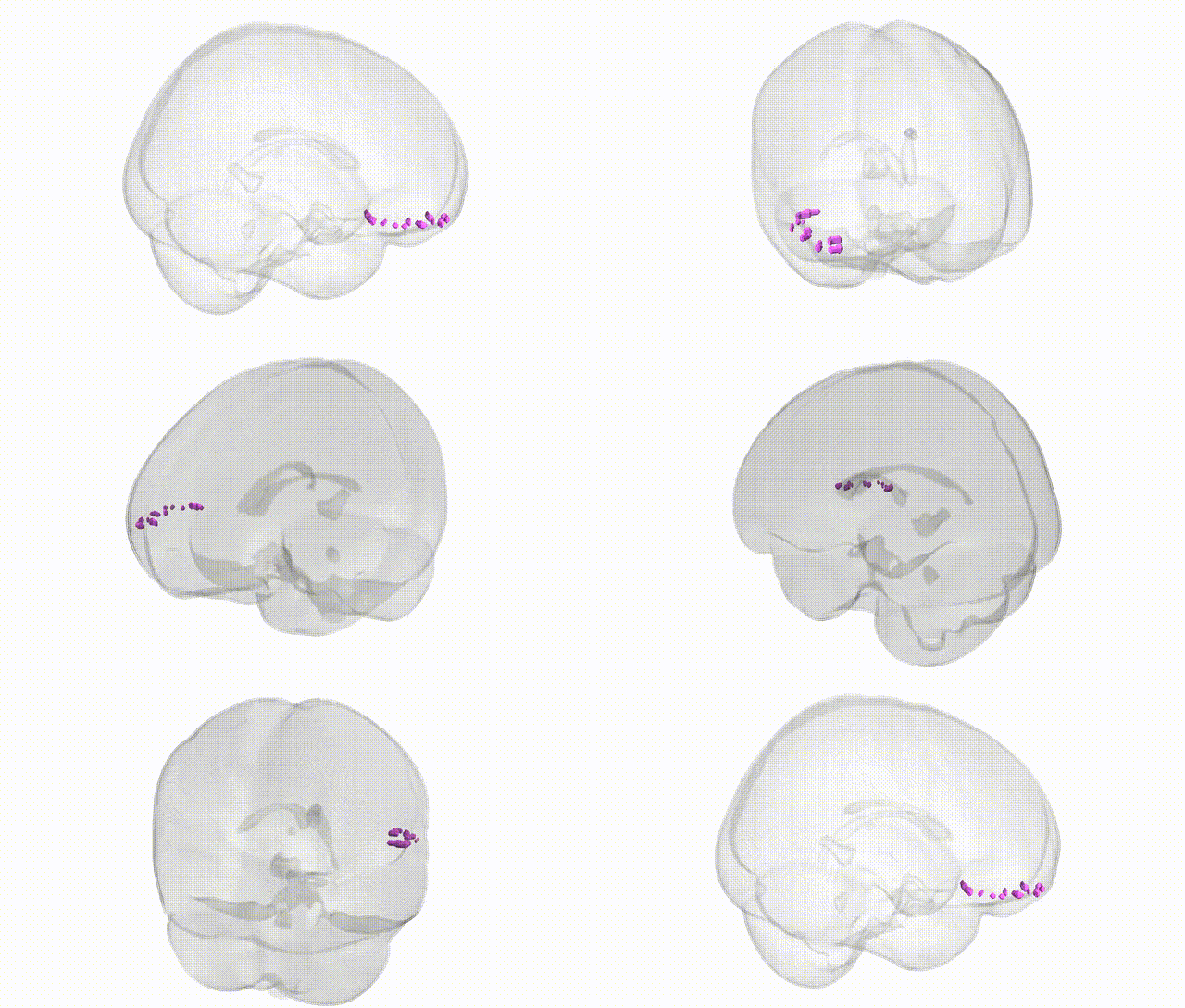
Full Quality Version: Download MP4
Wali Sidiqyar*, Gaurav Rudravaram*, Elyssa M. McMaster, Trent M. Schwartz, Adam M. Saunders, Kurt G. Schilling, Bennett A. Landman "Introducing SPINS: A Shared Public Visualization Library of Neuroanatomical Structures." Medical Imaging with Deep Learning- short paper
This resource is licensed under CC0 1.0 Universal (Public Domain).