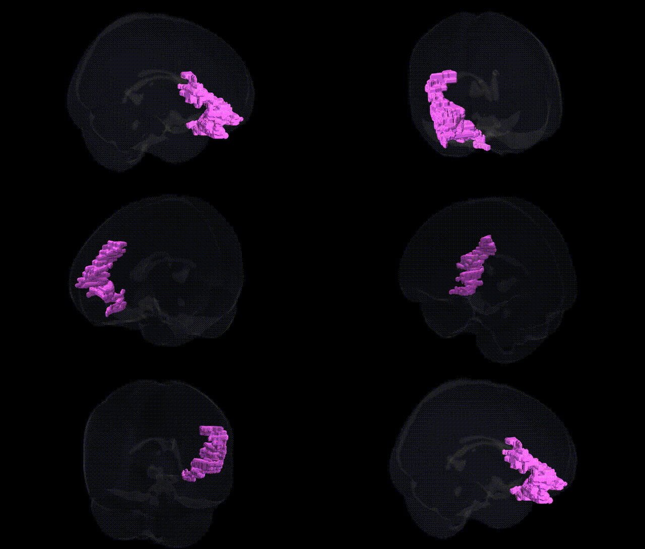
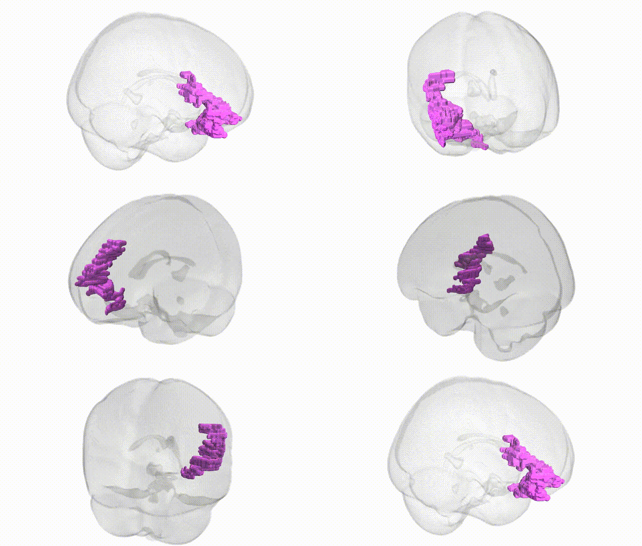
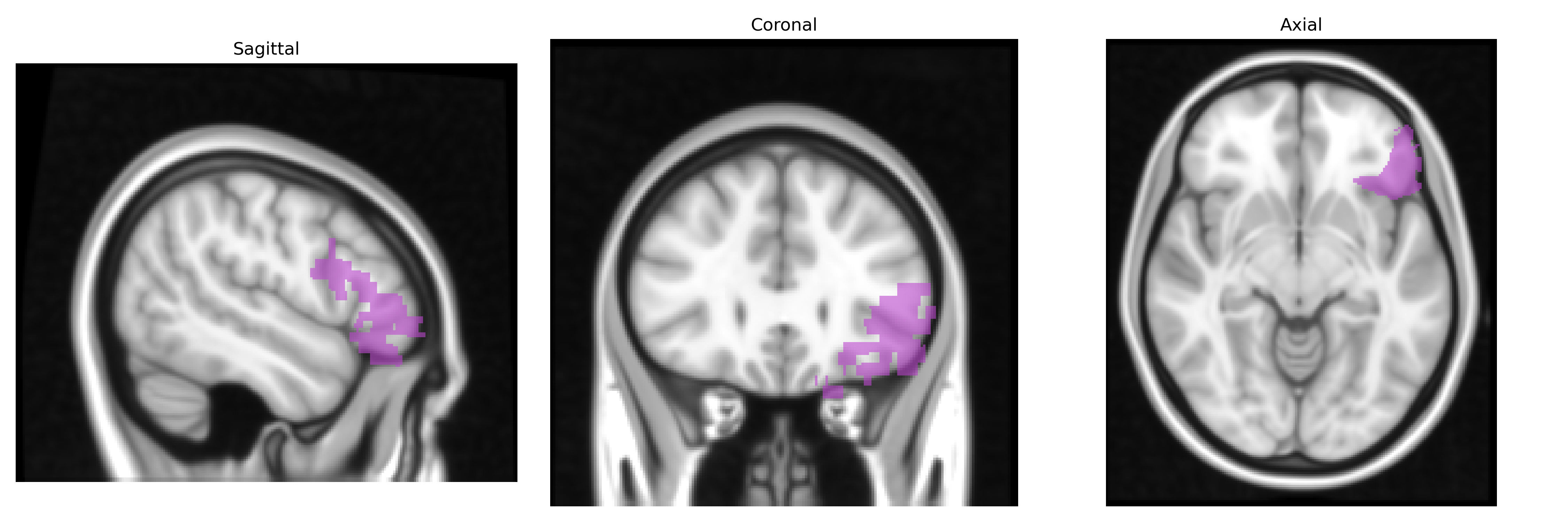
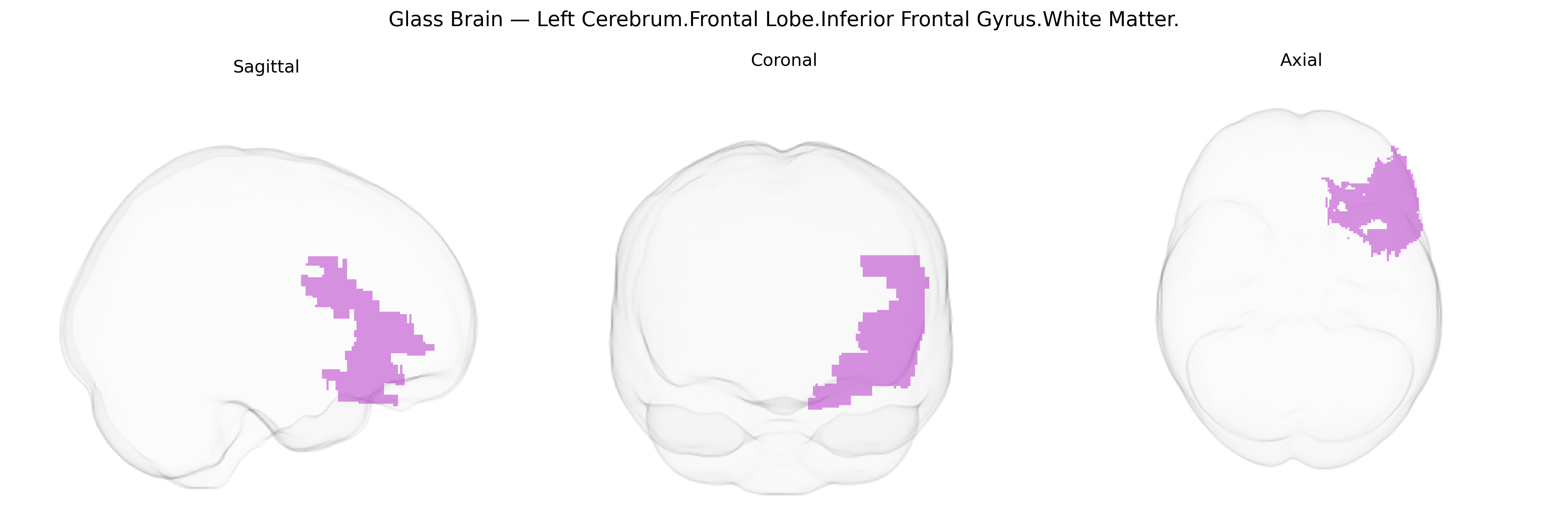
The bilateral Left Cerebrum.Frontal Lobe.Inferior Frontal Gyrus.White Matter corresponds to subcortical myelinated fiber pathways underlying the inferior frontal gyrus, a ventrolateral frontal region implicated in language, speech production, and aspects of cognitive control. These white matter tracts include short association fibers connecting adjacent frontal gyri and longer association pathways linking the inferior frontal cortex with temporal, parietal, and other frontal regions, thereby supporting networks involved in phonological and syntactic processing, verbal working memory, and response inhibition. In the left hemisphere, this white matter forms part of the broader perisylvian language circuitry, structurally coupling the inferior frontal gyrus with posterior language areas. There is no direct Wikipedia article for this specific white matter label; a related cortical region is the Inferior frontal gyrus.
The bilateral left inferior frontal gyrus (IFG) white matter, as defined in the Talairach 1 mm atlas, has been implicated in multiple genetic and GWAS-based associations primarily related to language, cognitive control, and psychiatric vulnerability. Polygenic influences on white matter microstructure in this region have been reported in large diffusion MRI GWAS, with variants in genes involved in axon guidance, myelination, and synaptic plasticity (e.g., NTRK3, CNTNAP2, and myelin-related loci) contributing to fractional anisotropy and connectivity measures linking the IFG to frontal–temporal language networks. Genetic variants associated with language and reading-related traits, including dyslexia and specific language impairment, have shown effects on IFG structure and connectivity, with loci in DCDC2, KIAA0319, and FOXP2-related pathways often highlighted in imaging-genetics work. In psychiatric genetics, schizophrenia, bipolar disorder, and major depression polygenic risk scores have been associated with altered IFG white matter integrity, while GWAS of impulsivity, attention-deficit/hyperactivity disorder, and externalizing traits have linked risk alleles in dopaminergic and glutamatergic genes to structural and functional connectivity between this region and other prefrontal and striatal areas. Additionally, autism spectrum disorder risk variants affecting synaptic and neurodevelopmental genes (e.g., SHANK3, NRXN1, and 16p11.2 CNVs) have been associated with atypical IFG morphology and white matter organization, consistent with the role of this region in social communication and language processing.
Overview generated by GPT-4o (2026).
Region ID: 189
Hemisphere: bilateral
Atlas: Talairach labels 1mm
Full Quality Version: Download MP4
Full Quality Version: Download MP4
Wali Sidiqyar*, Gaurav Rudravaram*, Elyssa M. McMaster, Trent M. Schwartz, Adam M. Saunders, Kurt G. Schilling, Bennett A. Landman "Introducing SPINS: A Shared Public Visualization Library of Neuroanatomical Structures." Medical Imaging with Deep Learning- short paper
This resource is licensed under CC0 1.0 Universal (Public Domain).