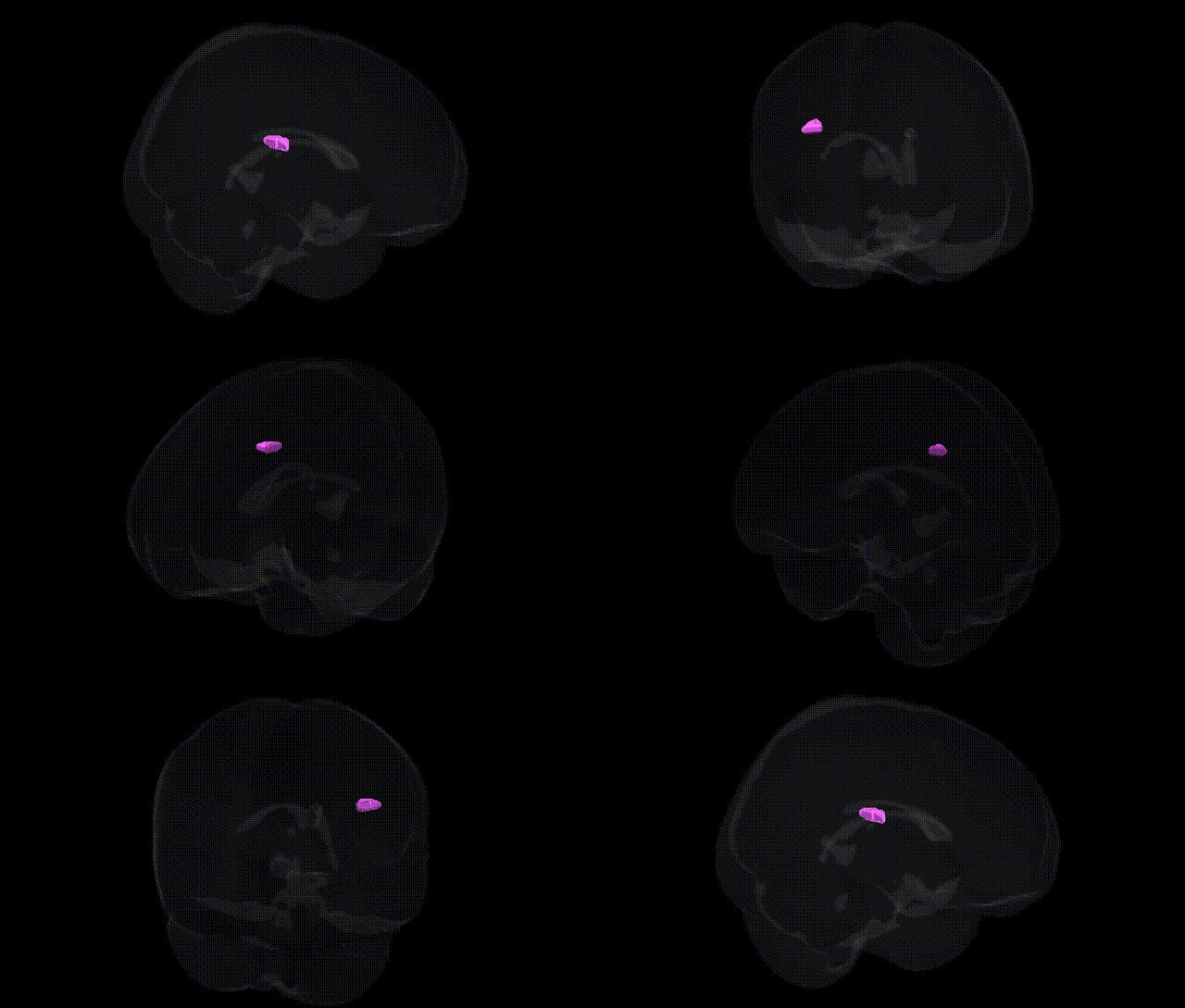
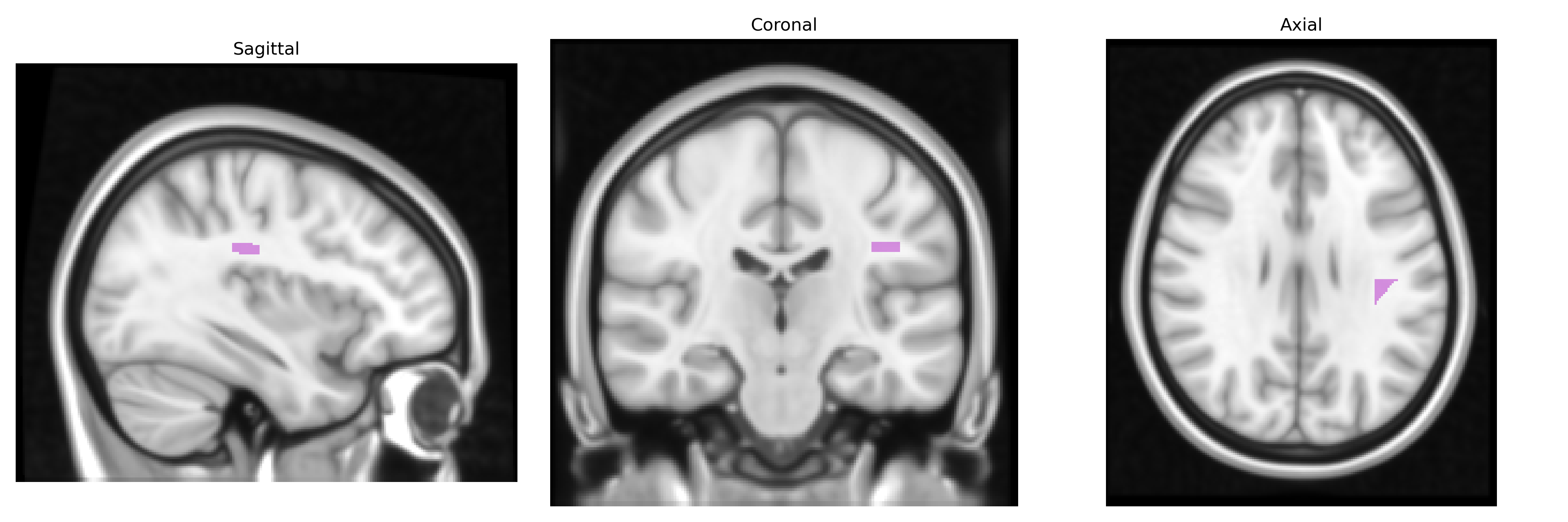
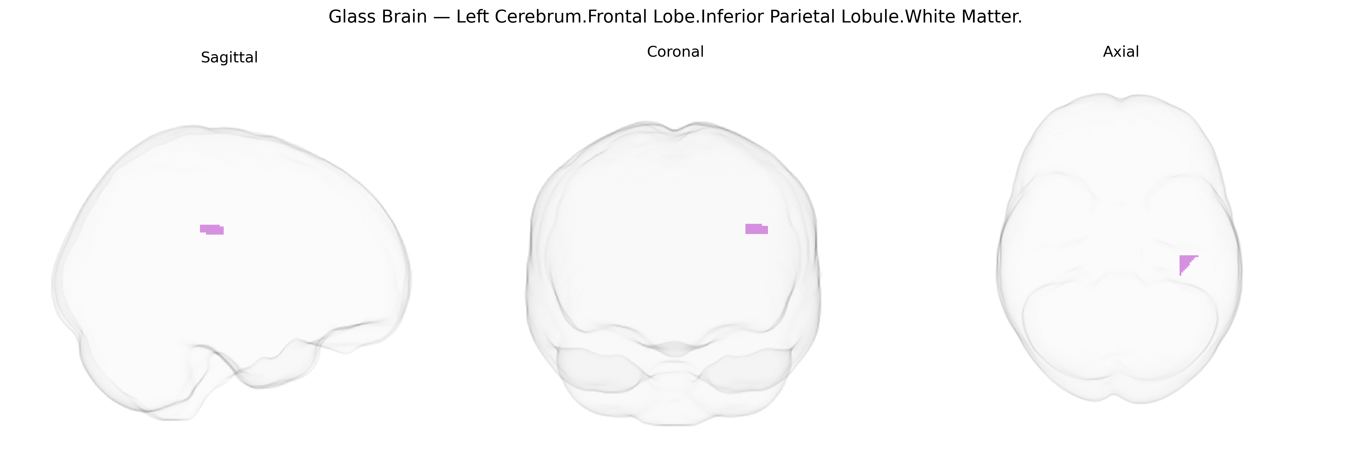
The bilateral Left Cerebrum.Frontal Lobe.Inferior Parietal Lobule.White Matter label in the Talairach 1 mm atlas refers to deep subcortical fibers underlying the inferior parietal lobule that extend anteriorly toward frontal lobe territories in the left hemisphere, reflecting long-range association and projection pathways rather than cortical gray matter per se. These white matter tracts are likely to include portions of major association bundles such as the superior longitudinal fasciculus and related fibers that interconnect inferior parietal regions with frontal cortical areas involved in language, attention, and higher-order sensorimotor integration, thereby supporting complex cognitive operations like phonological processing, working memory, and multimodal spatial analysis. There is no direct Wikipedia article for this specific white matter label; a related cortical region is the Inferior parietal lobule.
The bilateral left frontal–parietal white matter underlying the inferior parietal lobule, as defined in the Talairach 1 mm atlas, participates in frontoparietal association circuits whose microstructure shows robust heritability and multiple genetic associations in imaging-genetics and GWAS studies. Large consortia such as ENIGMA and UK Biobank have identified common variants influencing fractional anisotropy and other white-matter metrics in adjacent tracts (e.g., superior longitudinal fasciculus, arcuate fasciculus, and frontoparietal association fibers), with implicated loci including genes involved in axon guidance, myelination, and oligodendrocyte function (for example, variants near or within MAG, NTRK1/2, ERBB4, and genes in the neuregulin and semaphorin pathways). Polygenic overlap is reported between frontoparietal white-matter integrity and general cognitive ability (g), educational attainment, and working-memory performance, supporting a genetic contribution to individual differences in executive and attentional control mediated by this circuitry. GWAS of neuropsychiatric disorders have also linked risk alleles for schizophrenia, bipolar disorder, major depression, and ADHD to altered microstructure in frontoparietal white matter, with notable contributions from CACNA1C, ZNF804A, and several synaptic and calcium-channel genes, while autism spectrum disorder and dyslexia risk variants show associations with structural connectivity in parietal–frontal networks involved in language and social cognition. Collectively, these findings indicate that common genetic variation affecting neurodevelopment, synaptic signaling, and myelination contributes to structural and functional differences in left frontoparietal white matter, which in turn are implicated in a broad spectrum of cognitive traits and psychiatric or neurodevelopmental conditions.
Overview generated by GPT-4o (2026).
Region ID: 936
Hemisphere: bilateral
Atlas: Talairach labels 1mm
Full Quality Version: Download MP4
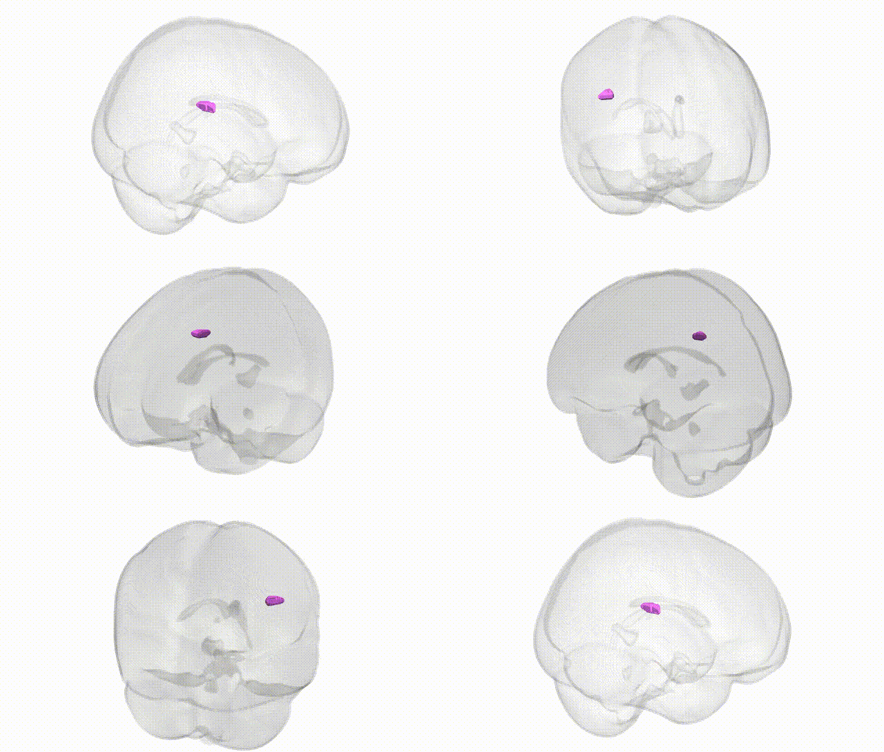
Full Quality Version: Download MP4
Wali Sidiqyar*, Gaurav Rudravaram*, Elyssa M. McMaster, Trent M. Schwartz, Adam M. Saunders, Kurt G. Schilling, Bennett A. Landman "Introducing SPINS: A Shared Public Visualization Library of Neuroanatomical Structures." Medical Imaging with Deep Learning- short paper
This resource is licensed under CC0 1.0 Universal (Public Domain).