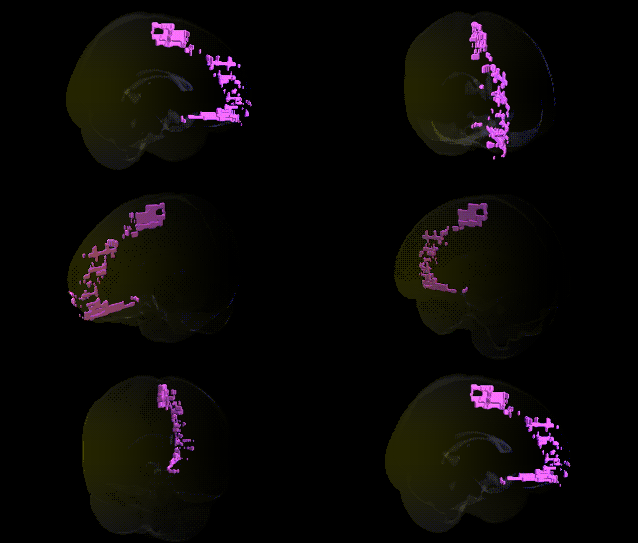
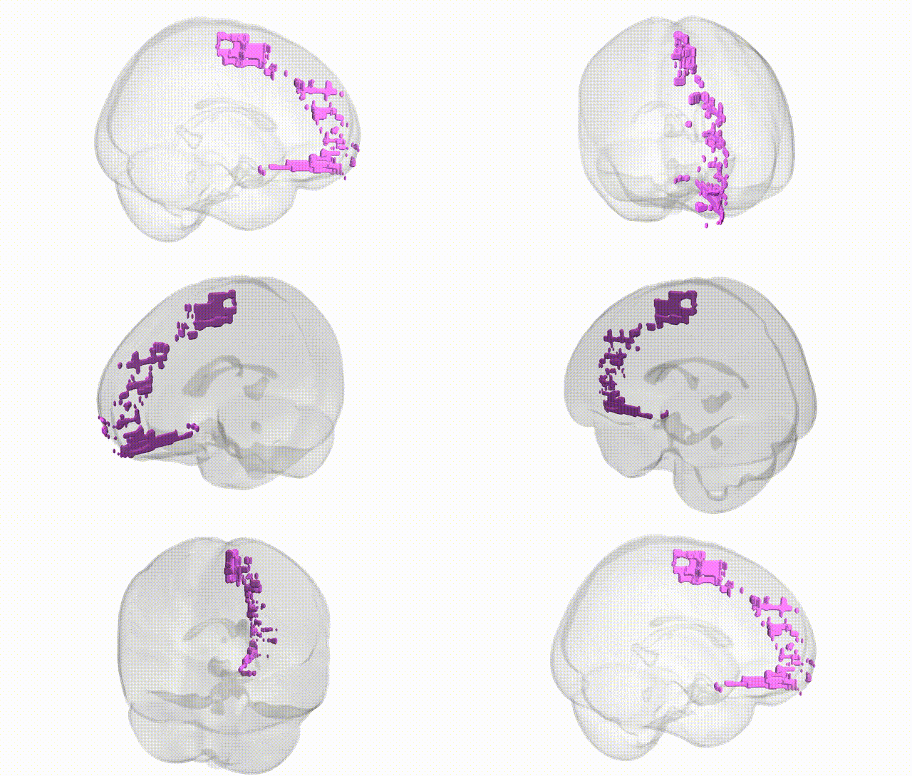
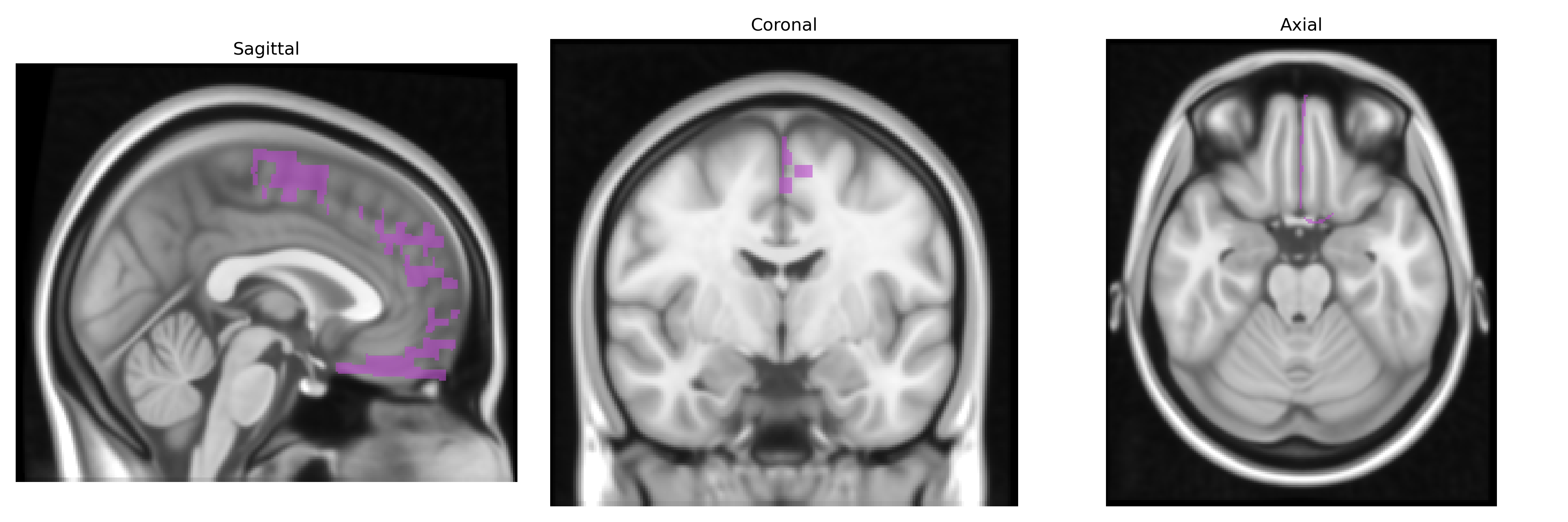
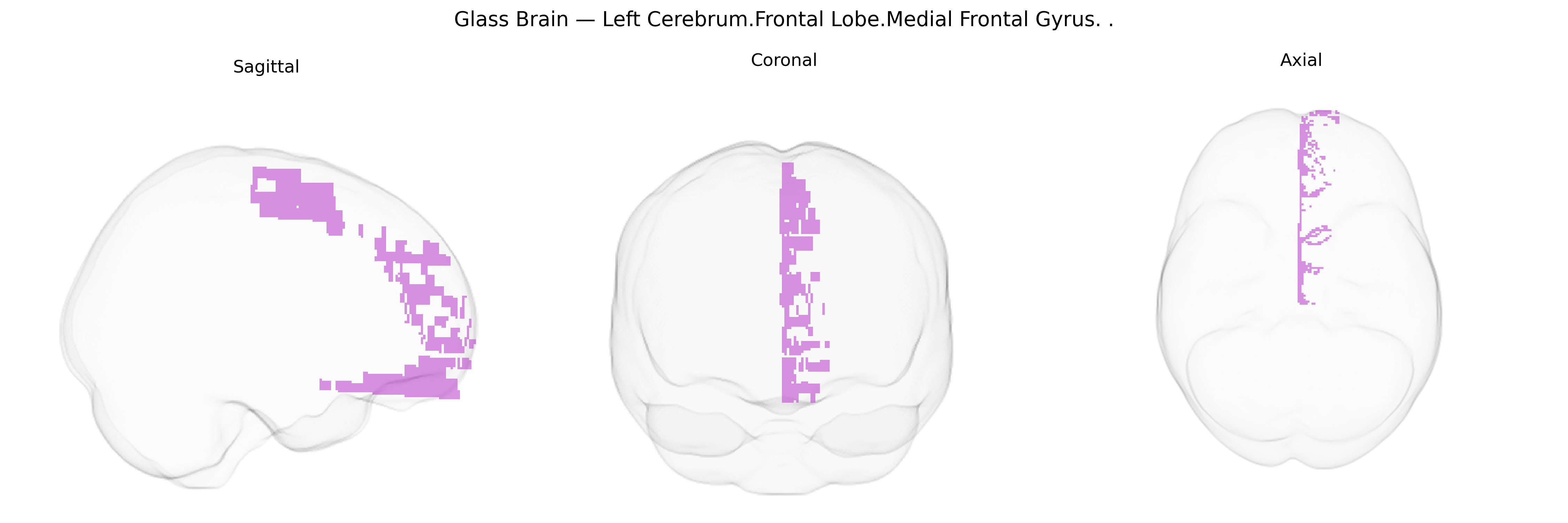
The bilateral Left Cerebrum.Frontal Lobe.Medial Frontal Gyrus, as defined in the Talairach 1 mm atlas, corresponds primarily to the medial portion of the superior and middle frontal gyri on the mesial surface of the frontal lobe, extending anteriorly from the supplementary motor area toward the frontal pole and superiorly along the interhemispheric fissure. This region is involved in higher-order executive functions, such as decision-making, attention control, motivation, and aspects of self-referential processing, and participates in motor planning, particularly in coordination with the supplementary and pre-supplementary motor areas. It forms part of large-scale networks including the default mode and frontoparietal control networks, with strong connectivity to the anterior cingulate cortex, prefrontal regions, and subcortical structures. There is no direct link for “Medial Frontal Gyrus”; a closely related structure is the Frontal Lobe.
The bilateral left medial frontal gyrus (Talairach 1 mm atlas: Left Cerebrum.Frontal Lobe.Medial Frontal Gyrus) has been implicated in multiple genetic and GWAS-based associations, largely via imaging genetics studies linking common variants to cortical thickness, surface area, and activation in medial frontal and adjacent anterior cingulate regions, with downstream links to psychiatric and cognitive traits. Variants in genes such as COMT, DRD2, BDNF, GRM3, DISC1, and NRG1 have been associated with structural and functional differences in medial frontal cortex, particularly in the context of schizophrenia, bipolar disorder, and major depressive disorder; large GWAS consortia (e.g., ENIGMA, UK Biobank) have identified polygenic influences on medial frontal morphology overlapping with risk loci for these disorders and for neuroticism, educational attainment, and general cognitive ability. Medial frontal regions encompassing the medial frontal gyrus show heritable variation in resting-state connectivity and task-related activation during cognitive control, reward processing, and error monitoring, with dopaminergic, glutamatergic, and GABAergic pathway genes frequently implicated. GWAS of obsessive–compulsive disorder, attention-deficit/hyperactivity disorder, and autism spectrum disorder have identified risk variants whose imaging correlates include altered medial frontal volume or activity, suggesting this area as a convergent site of genetic risk expression across multiple neurodevelopmental and psychiatric conditions. Additionally, polygenic scores for depression, anxiety, and substance use have been associated with altered medial prefrontal/medial frontal gyrus structure and function, supporting a role for this region as a key intermediate phenotype linking distributed genetic risk to affective and executive dysfunction.
Overview generated by GPT-4o (2026).
Region ID: 223
Hemisphere: bilateral
Atlas: Talairach labels 1mm
Full Quality Version: Download MP4
Full Quality Version: Download MP4
Wali Sidiqyar*, Gaurav Rudravaram*, Elyssa M. McMaster, Trent M. Schwartz, Adam M. Saunders, Kurt G. Schilling, Bennett A. Landman "Introducing SPINS: A Shared Public Visualization Library of Neuroanatomical Structures." Medical Imaging with Deep Learning- short paper
This resource is licensed under CC0 1.0 Universal (Public Domain).