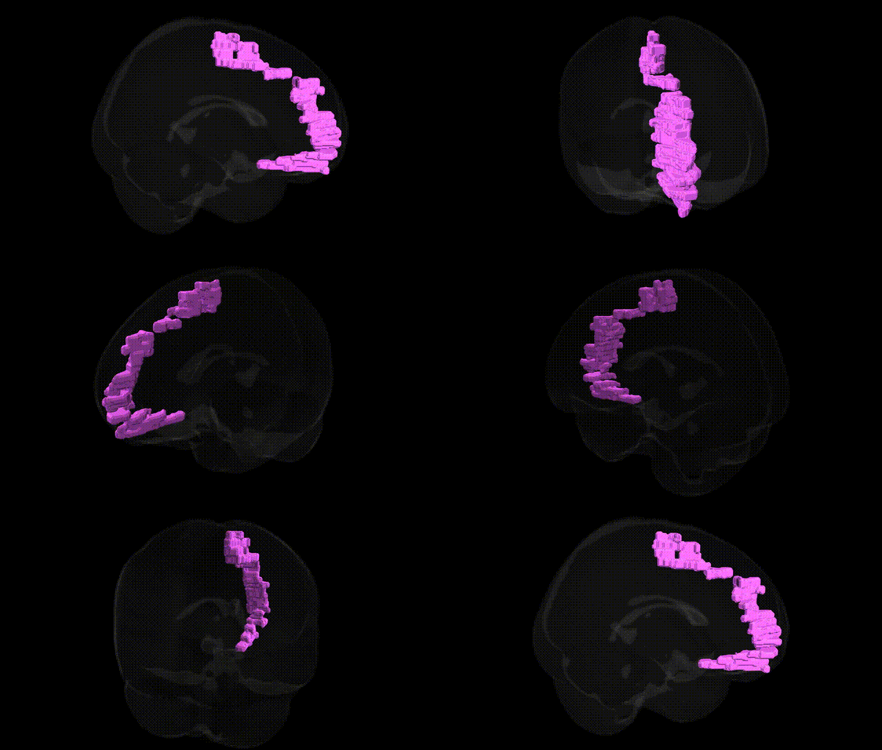
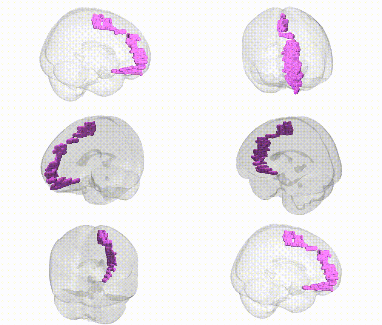
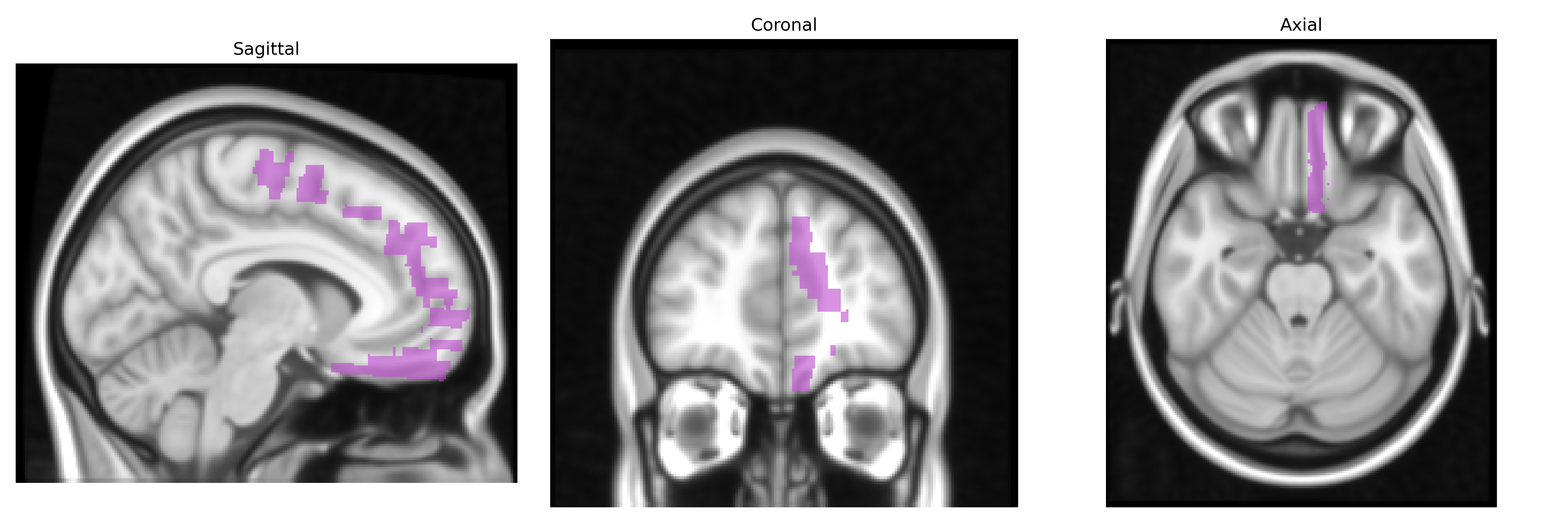
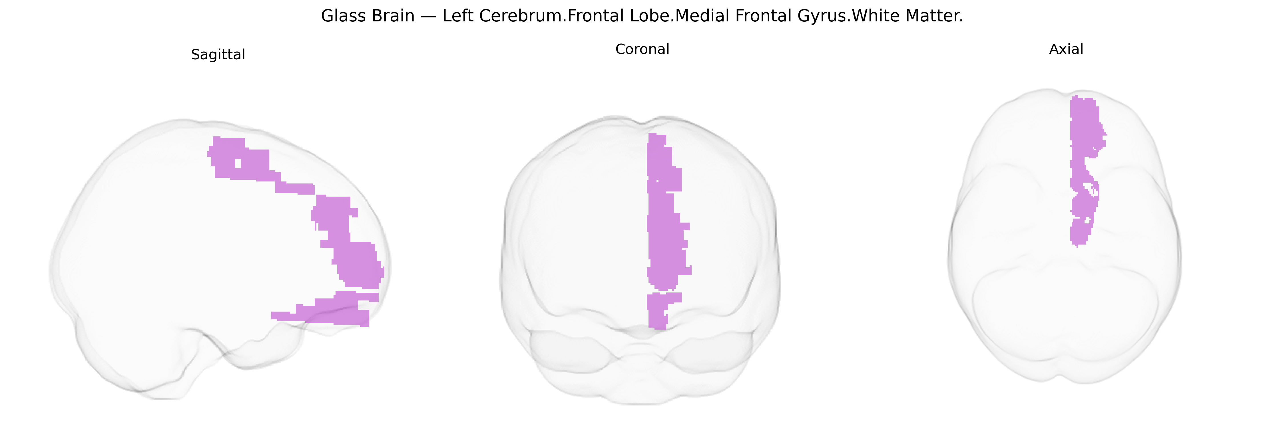
The bilateral Left Cerebrum.Frontal Lobe.Medial Frontal Gyrus.White Matter corresponds to the subcortical myelinated fiber pathways underlying the medial frontal gyrus in the frontal lobe, extending along the medial surface of the cerebral hemisphere anterior to the supplementary motor and cingulate regions. This white matter compartment contains association fibers connecting medial prefrontal, orbitofrontal, and dorsolateral frontal cortices, as well as projections to and from limbic, cingulate, and subcortical structures, supporting higher-order executive, motivational, and attentional processes. It is situated superior to the corpus callosum and anterior cingulate cortex and is part of the broader medial prefrontal white matter system rather than a distinct gross anatomical tract. There is no direct link for this specific white matter label; a closely related cortical region is the Medial frontal gyrus.
The medial frontal gyrus white matter of the left frontal lobe, as defined in Talairach 1 mm space, lies within a broader medial prefrontal/anterior cingulate–adjacent network whose microstructure and volume show modest but consistent heritability and multiple genome-wide associations with neuropsychiatric and cognitive traits. Large GWAS of diffusion MRI (e.g., UK Biobank–based studies) have identified common variants in genes involved in axon guidance, myelination, and synaptic function—such as NRG1/NRG3, CNTN4/6, MAG, and multiple loci near oligodendrocyte-related genes—associated with fractional anisotropy and mean diffusivity in medial frontal and cingulum white matter, which anatomically traverse or border the medial frontal gyrus. Polygenic risk scores for schizophrenia, bipolar disorder, and major depressive disorder are associated with reduced integrity or altered volume in medial prefrontal and adjacent cingulum white matter, while risk loci in CACNA1C, ZNF804A, and other synaptic and calcium-channel genes track with these alterations. Additional GWAS of cortical morphology implicate loci near genes such as MIR137, TCF4, and multiple glutamatergic and GABAergic pathway genes in variation of medial prefrontal thickness and surface area, which are structurally continuous with the medial frontal gyrus and supported by its underlying white matter. Medial frontal white-matter metrics in this region are further associated, at the genetic level, with traits including general cognitive ability, educational attainment, neuroticism, and risk-related decision-making, reflecting the region’s role in higher-order control and affective regulation, although atlas-level, Talairach-specific labels are rarely the direct unit of analysis in these genome-wide studies.
Overview generated by GPT-4o (2026).
Region ID: 230
Hemisphere: bilateral
Atlas: Talairach labels 1mm
Full Quality Version: Download MP4
Full Quality Version: Download MP4
Wali Sidiqyar*, Gaurav Rudravaram*, Elyssa M. McMaster, Trent M. Schwartz, Adam M. Saunders, Kurt G. Schilling, Bennett A. Landman "Introducing SPINS: A Shared Public Visualization Library of Neuroanatomical Structures." Medical Imaging with Deep Learning- short paper
This resource is licensed under CC0 1.0 Universal (Public Domain).