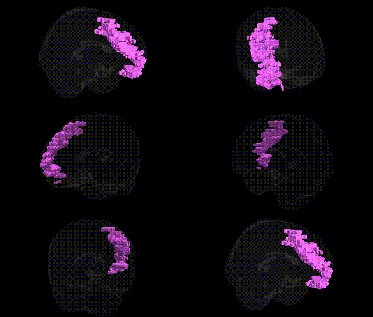
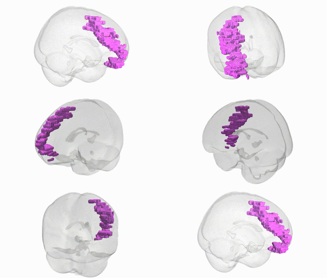
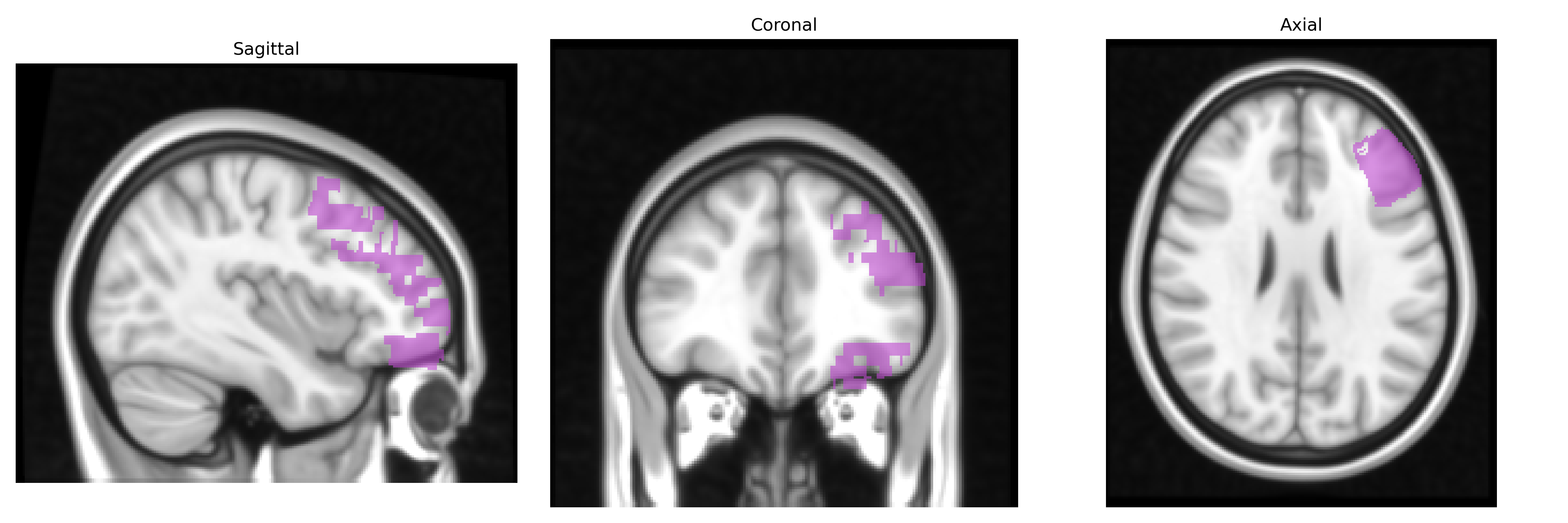
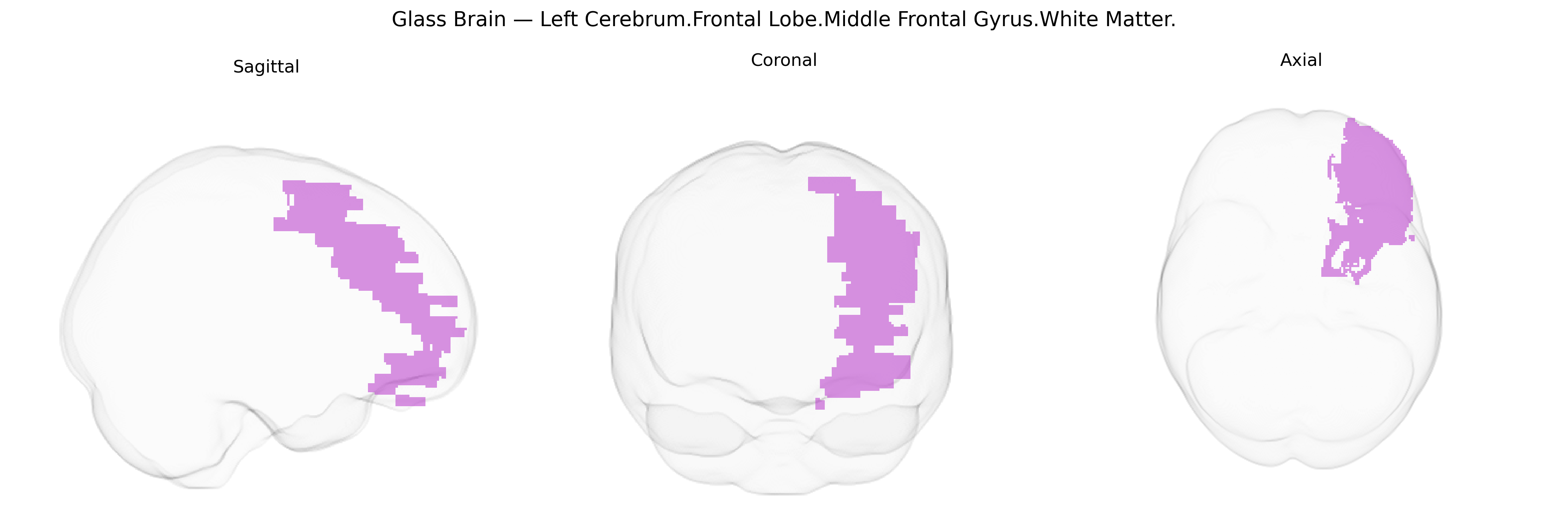
The bilateral Left Cerebrum.Frontal Lobe.Middle Frontal Gyrus.White Matter corresponds to the subcortical fiber systems underlying the middle frontal gyrus, a key portion of the dorsolateral prefrontal cortex involved in higher-order cognitive functions such as working memory, executive control, planning, and attentional regulation. This white matter contains association fibers that interconnect adjacent and distant frontal cortical regions, as well as pathways linking frontal areas to parietal, temporal, and subcortical structures, thereby supporting integrative processing required for complex behavior and decision-making. It contributes to the structural connectivity of prefrontal networks implicated in cognitive flexibility, goal-directed behavior, and modulation of motor and premotor systems. There is no direct link for this exact white matter subdivision; a related cortical structure is the Middle frontal gyrus.
Genetic associations with the bilateral Left Cerebrum.Frontal Lobe.Middle Frontal Gyrus.White Matter (approximately overlapping with dorsolateral prefrontal and adjacent frontal white matter tracts in the Talairach 1 mm atlas) largely arise from imaging–genetics and GWAS of brain structure and connectivity, rather than direct locus-specific studies. Large consortia such as ENIGMA and UK Biobank have identified variants in genes involved in neurodevelopment, axon guidance, and myelination (for example, NRG1/3, CNTNAP2, MAG, and other oligodendrocyte-related genes) that influence frontal lobe white matter volume, cortical thickness, and diffusion metrics in this region or closely overlapping tracts (e.g., superior longitudinal fasciculus, anterior corona radiata). Polygenic risk for schizophrenia, bipolar disorder, and major depressive disorder is associated with altered middle frontal white matter microstructure and volume, suggesting shared genetic architectures between psychiatric risk loci (often in synaptic, calcium-channel, and neurodevelopmental genes) and structural integrity of this area. Variants linked to cognitive ability and educational attainment (notably in loci regulating synaptic plasticity and cortical development) show associations with frontal white matter measures, aligning with the role of the middle frontal gyrus in working memory and executive function. Additional GWAS of white matter hyperintensities, small vessel disease, and general brain aging identify vascular and inflammatory genes (such as those in blood–brain barrier integrity and lipid metabolism pathways) that contribute to lesion burden and microstructural changes in frontal white matter, which includes the middle frontal region. Overall, the genetic landscape connected to this region reflects a convergence of neurodevelopmental, myelination, vascular, and psychiatric risk pathways rather than a single, region-specific set of loci.
Overview generated by GPT-4o (2026).
Region ID: 195
Hemisphere: bilateral
Atlas: Talairach labels 1mm
Full Quality Version: Download MP4
Full Quality Version: Download MP4
Wali Sidiqyar*, Gaurav Rudravaram*, Elyssa M. McMaster, Trent M. Schwartz, Adam M. Saunders, Kurt G. Schilling, Bennett A. Landman "Introducing SPINS: A Shared Public Visualization Library of Neuroanatomical Structures." Medical Imaging with Deep Learning- short paper
This resource is licensed under CC0 1.0 Universal (Public Domain).