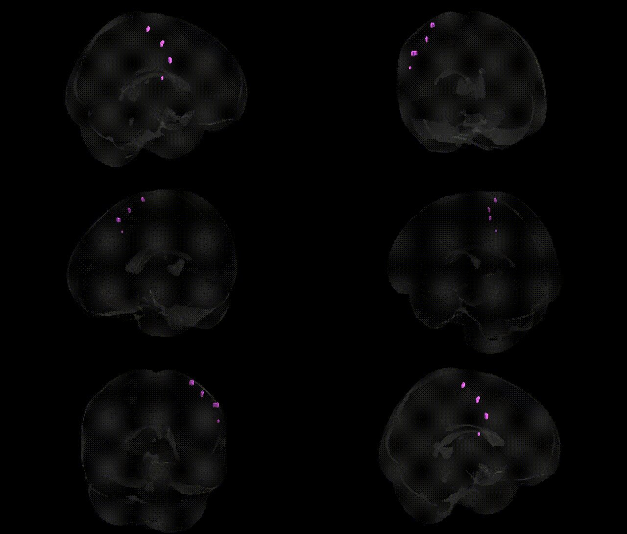
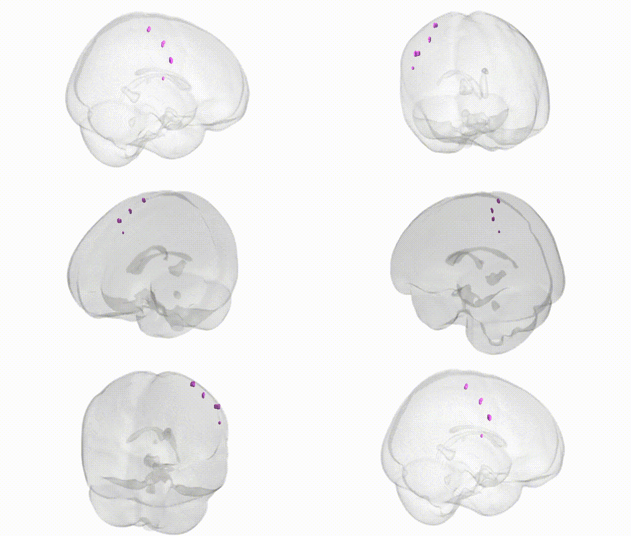
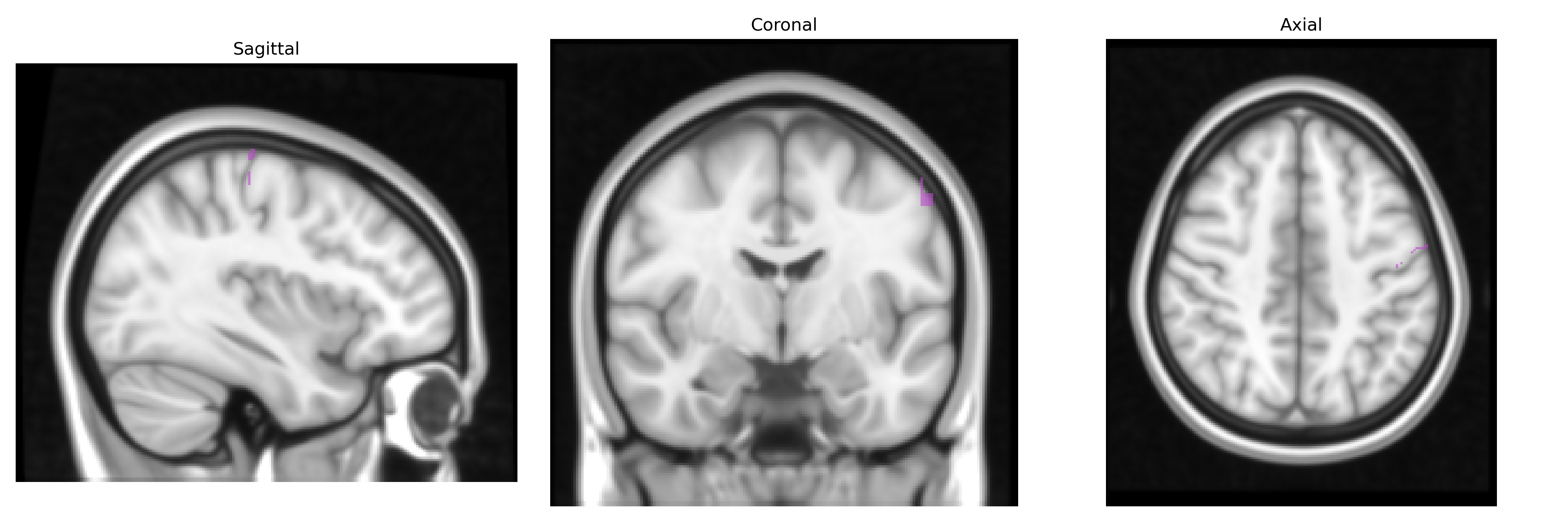
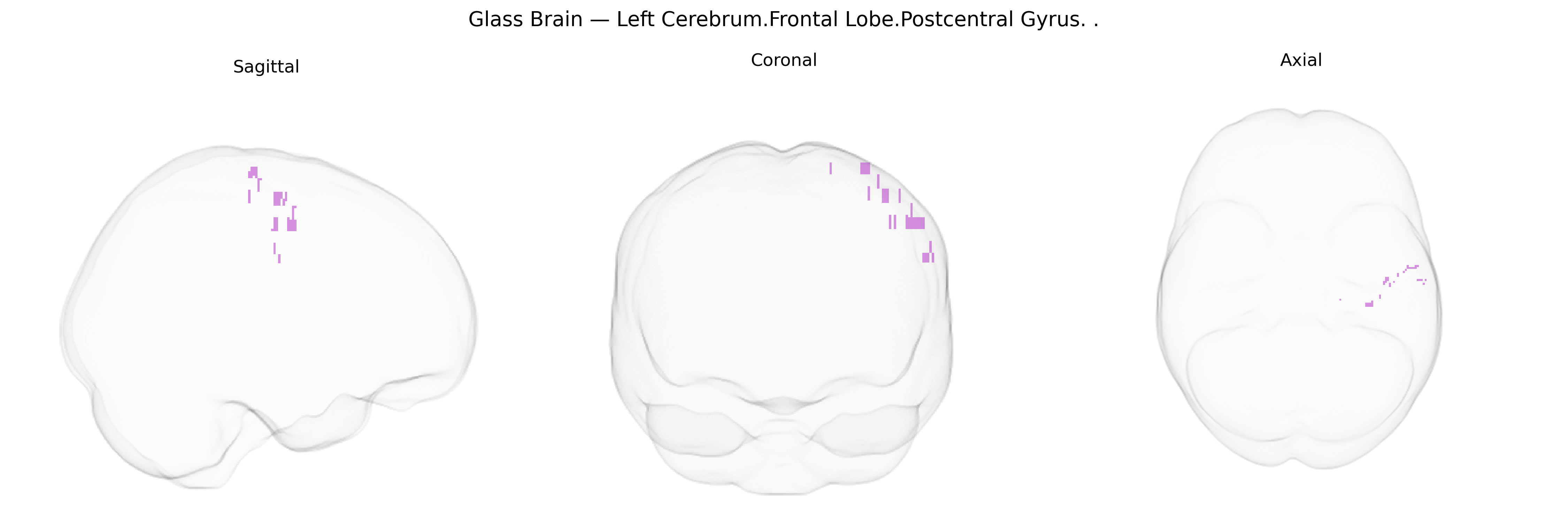
The bilateral Left Cerebrum.Frontal Lobe.Postcentral Gyrus label in the Talairach 1 mm Atlas most likely reflects a border or misassigned region near the central sulcus, as the postcentral gyrus is classically part of the parietal lobe rather than the frontal lobe. The postcentral gyrus corresponds to the primary somatosensory cortex (Brodmann areas 3, 1, and 2), which receives and processes tactile, proprioceptive, and nociceptive information from the contralateral side of the body via thalamocortical projections. Its organization is somatotopic, forming a sensory homunculus in which discrete cortical territories represent specific body regions, with medial portions devoted to lower limbs and more lateral regions representing trunk, upper limb, and face. This cortex plays a central role in conscious perception of touch, vibration, joint position, and pain, and contributes to sensorimotor integration by projecting to motor and premotor regions, thereby influencing movement planning and execution. Postcentral gyrus
The bilateral left frontal/postcentral region in the Talairach 1 mm atlas corresponds primarily to primary motor and adjacent premotor/somatosensory cortex, for which genetic associations have been inferred largely through GWAS of cortical thickness, surface area, and gyrification, as well as imaging–genetics and lesion–mapping studies of motor and somatosensory function. Variants in genes involved in neuronal development and synaptic function—such as MAPT, BDNF, HRNBP3, FOXP2-related networks, and polygenic scores for neurodevelopmental traits—have been linked to structural variation in nearby precentral/postcentral and frontal sensorimotor regions, while large-scale ENIGMA and UK Biobank analyses have identified multiple loci (e.g., on chromosomes 3p, 6q, 8q, 17q) associated with thickness and surface area in precentral/postcentral and superior frontal areas that spatially overlap or neighbor this label. Clinically, genetic risk for amyotrophic lateral sclerosis (e.g., C9orf72 repeat expansions and other ALS loci), primary lateral sclerosis, and certain hereditary spastic paraplegias shows preferential atrophy and microstructural changes in the motor strip and adjacent frontal cortex, while polygenic risk scores for schizophrenia, ADHD, and autism have been associated with altered morphology and activation in frontal motor and somatosensory regions. In addition, GWAS of handedness and motor skill learning implicate loci involved in axon guidance, synaptic plasticity, and cortical patterning that influence asymmetry and connectivity of frontal and parietal motor networks encompassing this area.
Overview generated by GPT-4o (2026).
Region ID: 979
Hemisphere: bilateral
Atlas: Talairach labels 1mm
Full Quality Version: Download MP4
Full Quality Version: Download MP4
Wali Sidiqyar*, Gaurav Rudravaram*, Elyssa M. McMaster, Trent M. Schwartz, Adam M. Saunders, Kurt G. Schilling, Bennett A. Landman "Introducing SPINS: A Shared Public Visualization Library of Neuroanatomical Structures." Medical Imaging with Deep Learning- short paper
This resource is licensed under CC0 1.0 Universal (Public Domain).