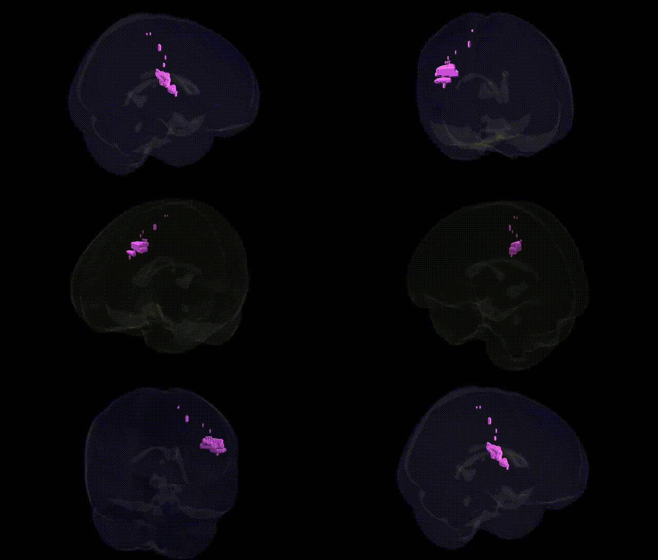
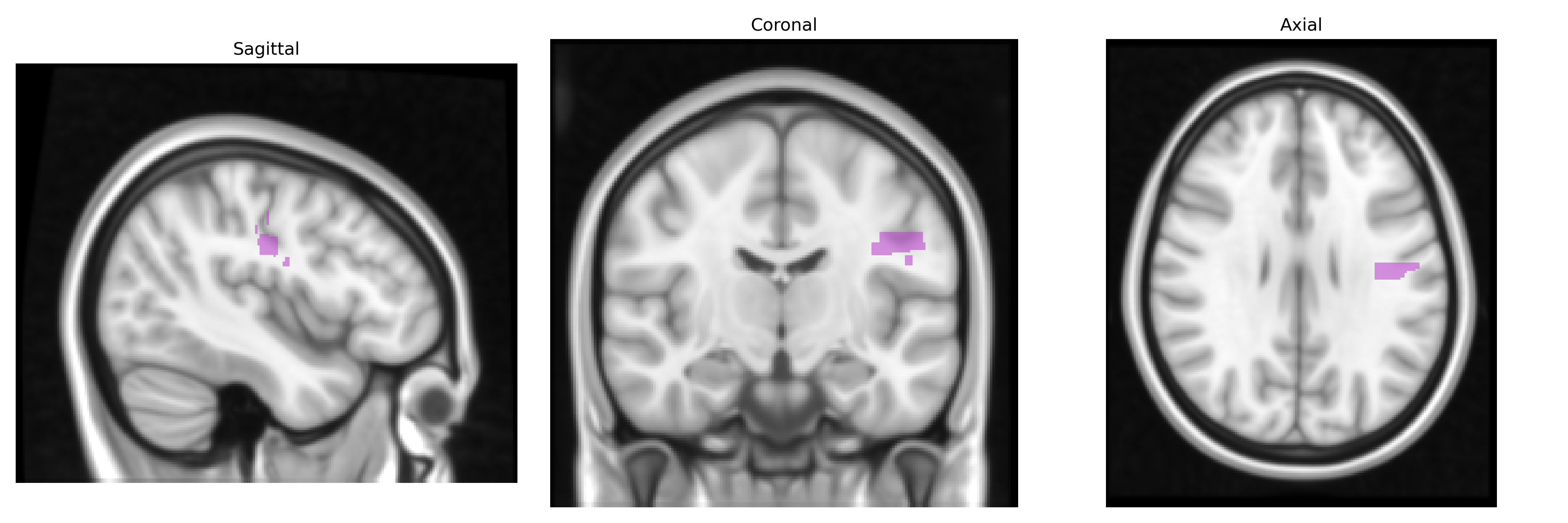
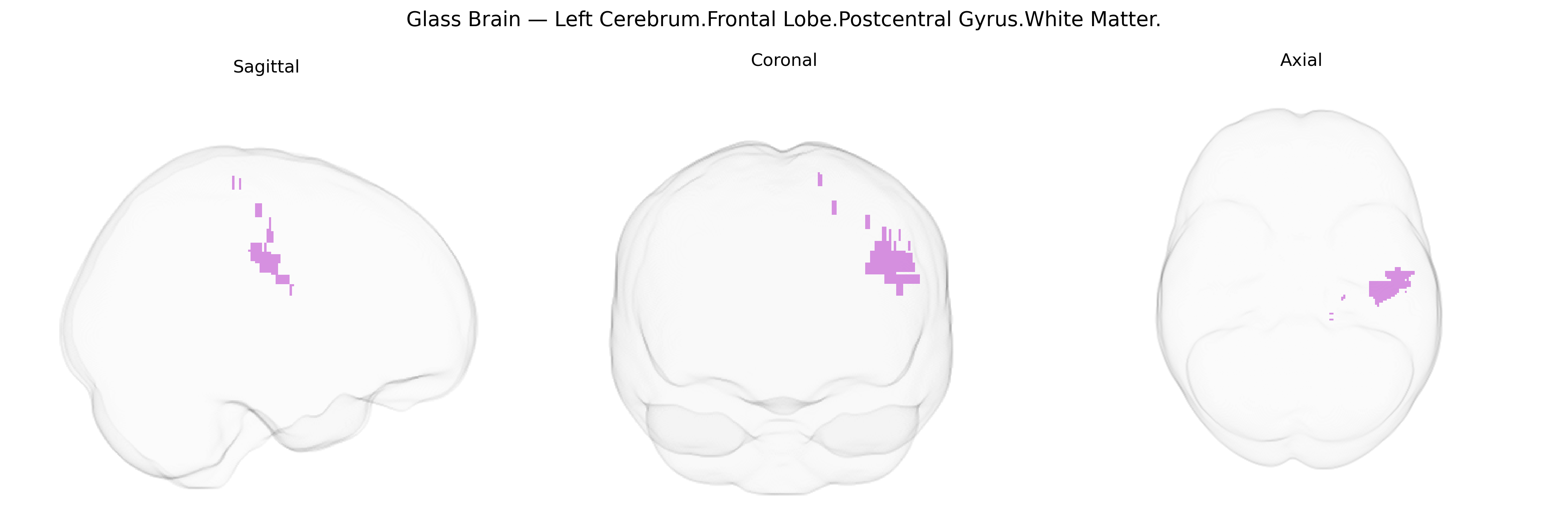
The “bilateral Left Cerebrum.Frontal Lobe.Postcentral Gyrus.White Matter” label from the Talairach 1 mm atlas appears to reflect a misregistration or naming inconsistency, as the postcentral gyrus is classically part of the parietal lobe rather than the frontal lobe. Anatomically, white matter deep to the postcentral gyrus comprises ascending thalamocortical fibers (primarily from the ventral posterior nucleus of the thalamus) and descending corticocortical and corticofugal fibers that interconnect primary somatosensory cortex with premotor, motor, and posterior parietal areas, as well as subcortical structures. Functionally, this white matter supports the rapid transmission and integration of tactile, proprioceptive, and nociceptive information required for somatosensory perception, sensorimotor integration, and fine motor control. There is no direct Wikipedia article for this specific white-matter label; a closely related structure is the Postcentral gyrus.
Genetic associations involving bilateral frontal/postcentral white-matter regions corresponding to the “Left Cerebrum.Frontal Lobe.Postcentral Gyrus.White Matter” label in the Talairach 1 mm atlas largely emerge from imaging–genetics and GWAS of white-matter microstructure (e.g., FA, MD) in corticospinal and frontoparietal association tracts that traverse this area, such as the superior longitudinal fasciculus and adjacent sensorimotor pathways. Large-scale studies have identified variants in genes involved in axon guidance, myelination, and neuronal development (for example, NTRK1/2, CNTN4, MAG, and other oligodendrocyte- and myelin-related loci) as influencing white-matter integrity in frontal and postcentral regions, often overlapping with risk loci for neurodevelopmental and psychiatric conditions, including schizophrenia, ADHD, and autism spectrum disorder, where altered sensorimotor and frontal white matter is a consistent imaging finding. Polygenic scores for cognitive performance, educational attainment, and general intelligence have been associated with microstructural variation in frontoparietal white matter, suggesting that common genetic variation affecting axonal organization and myelin impacts both structural connectivity in this region and higher-order cognitive traits. Additionally, GWAS of stroke, small-vessel disease, and leukoaraiosis have implicated vascular and endothelial genes (e.g., COL4A2, HTRA1, NOTCH3-related pathways) in white-matter hyperintensities that frequently involve pericentral and frontal white matter, linking genetic susceptibility for microvascular pathology to structural alterations in this Talairach-defined region.
Overview generated by GPT-4o (2026).
Region ID: 833
Hemisphere: bilateral
Atlas: Talairach labels 1mm
Full Quality Version: Download MP4
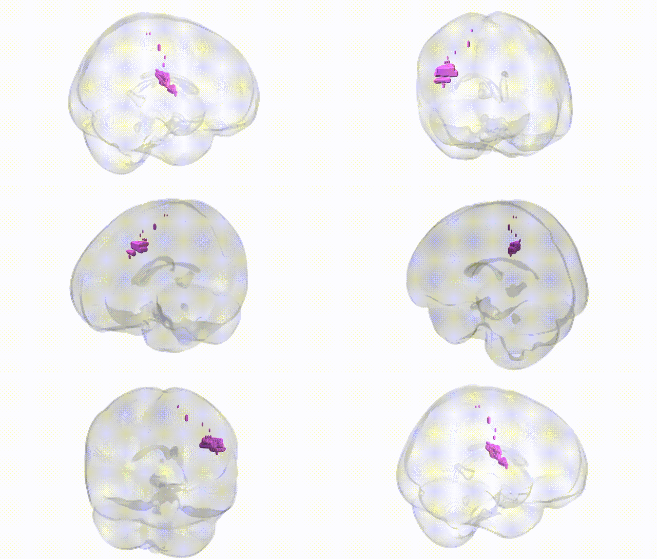
Full Quality Version: Download MP4
Wali Sidiqyar*, Gaurav Rudravaram*, Elyssa M. McMaster, Trent M. Schwartz, Adam M. Saunders, Kurt G. Schilling, Bennett A. Landman "Introducing SPINS: A Shared Public Visualization Library of Neuroanatomical Structures." Medical Imaging with Deep Learning- short paper
This resource is licensed under CC0 1.0 Universal (Public Domain).