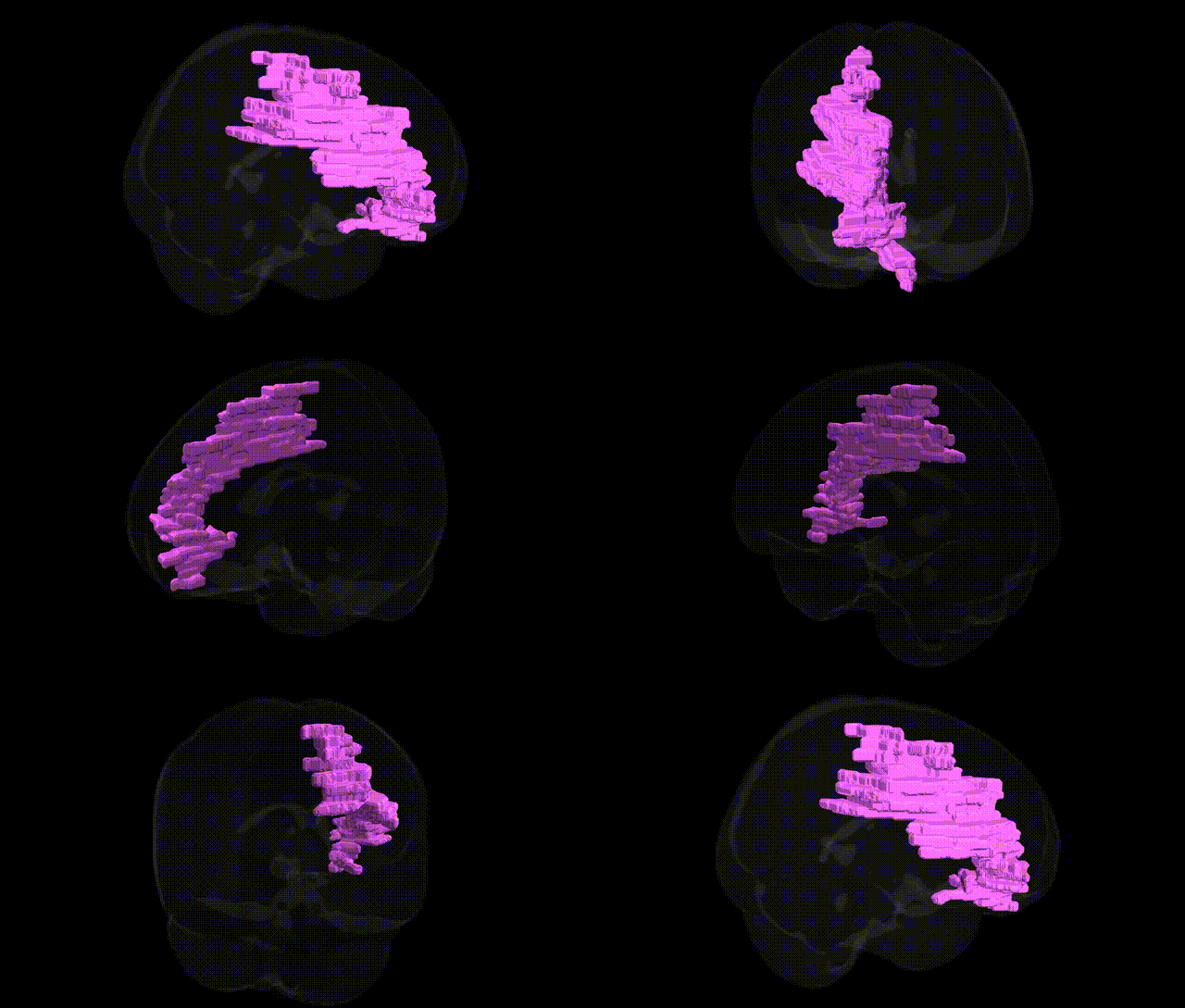
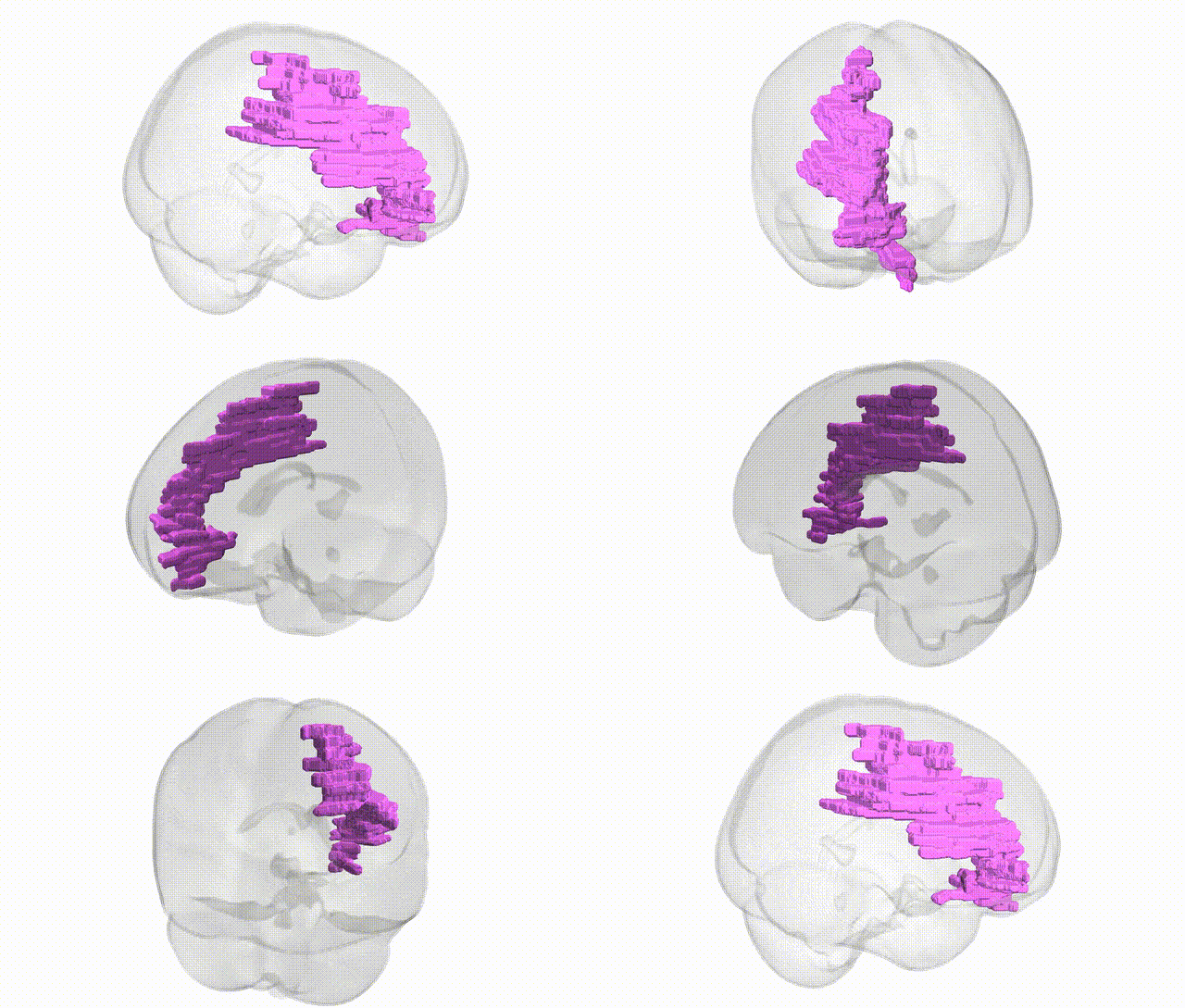
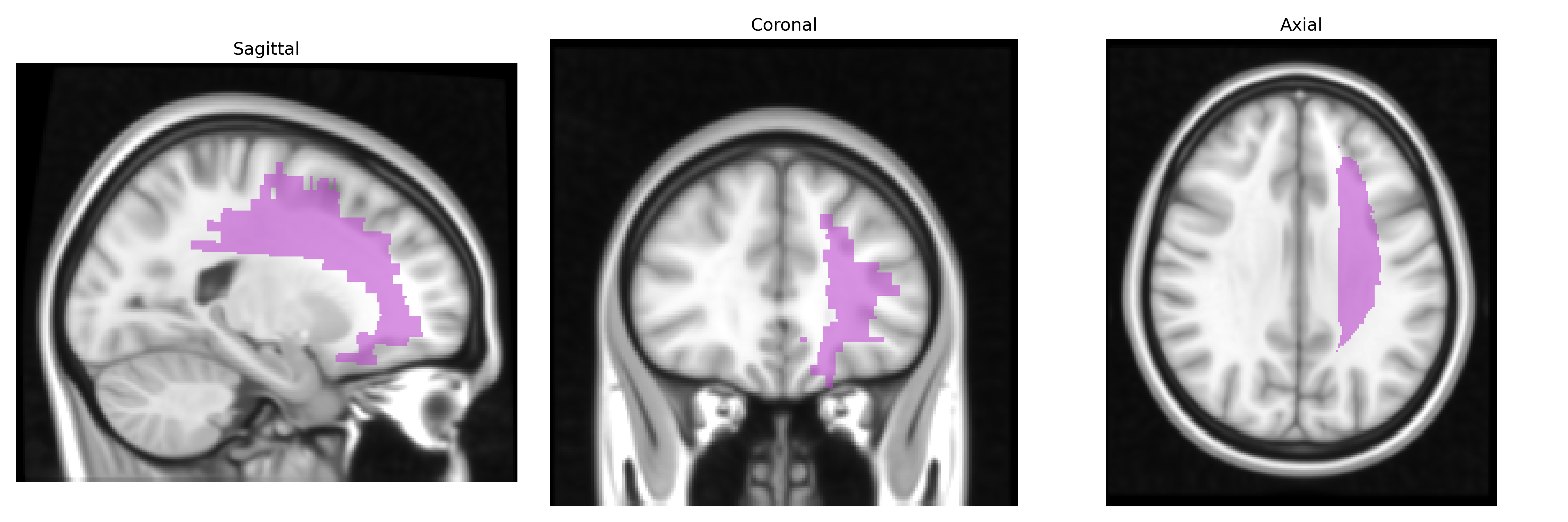
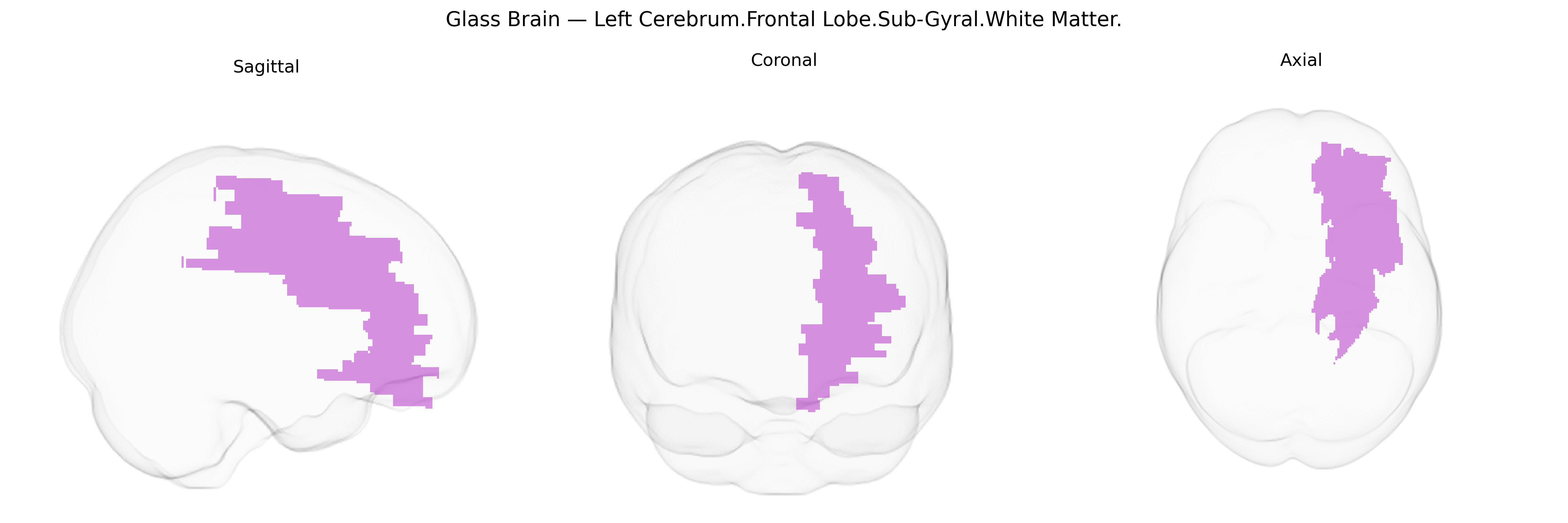
The bilateral Left Cerebrum.Frontal Lobe.Sub-Gyral.White Matter refers to deep frontal lobe white matter lying beneath the cortical gyri of the left frontal lobe, comprising bundles of myelinated axons that interconnect frontal cortical areas with each other and with distant regions such as the parietal, temporal, occipital lobes, basal ganglia, thalamus, and brainstem. This subgyral white matter includes portions of major association, commissural, and projection pathways that support executive functions, working memory, attention, language production, motor planning, and aspects of social and emotional processing by enabling efficient communication within frontal networks and between frontal cortex and other brain systems. As a structural rather than cytoarchitectonically defined area, it is functionally heterogeneous, with specific fiber tracts contributing to distinct cognitive and motor operations depending on their cortical and subcortical connections. There is no direct link for this exact Talairach label; see the related structure Frontal lobe.
The bilateral left frontal sub-gyral white matter (as delineated in Talairach 1 mm space) has been implicated in genetic studies primarily through its role in major white matter tracts underlying dorsolateral prefrontal and premotor cortices, with associations emerging from GWAS of brain structure, connectivity, and neuropsychiatric disorders. Variants in genes affecting myelination and axonal integrity (for example, oligodendrocyte- and myelin-related genes such as those within the major histocompatibility complex, NRG1/ErbB signaling, and other neurodevelopmental pathways) have been linked to fractional anisotropy and white matter volume in frontal regions that include sub-gyral zones. Large-scale imaging-genetics consortia (e.g., ENIGMA, UK Biobank imaging GWAS) report heritable variation in frontal white matter microstructure, with loci near genes involved in axon guidance, cytoskeletal dynamics, and synaptic development associated with diffusion MRI measures in frontal lobe tracts such as the superior longitudinal fasciculus and anterior corona radiata, which traverse sub-gyral white matter. Clinically, genetic risk for schizophrenia, bipolar disorder, and major depression—indexed by polygenic risk scores and disorder-specific loci—has been associated with reduced integrity or altered volume of frontal white matter, while common variants linked to cognitive ability, educational attainment, and ADHD show convergent effects on prefrontal white matter organization. Further, susceptibility genes for small-vessel disease and ischemic stroke (e.g., in NOTCH3 and other vascular-related loci) contribute to frontal white matter hyperintensities and microstructural disruption, underscoring a genetic contribution to the vulnerability of this region in both neuropsychiatric and cerebrovascular phenotypes, even though few GWAS target the Talairach-defined bilateral left frontal sub-gyral white matter as a standalone region.
Overview generated by GPT-4o (2026).
Region ID: 193
Hemisphere: bilateral
Atlas: Talairach labels 1mm
Full Quality Version: Download MP4
Full Quality Version: Download MP4
Wali Sidiqyar*, Gaurav Rudravaram*, Elyssa M. McMaster, Trent M. Schwartz, Adam M. Saunders, Kurt G. Schilling, Bennett A. Landman "Introducing SPINS: A Shared Public Visualization Library of Neuroanatomical Structures." Medical Imaging with Deep Learning- short paper
This resource is licensed under CC0 1.0 Universal (Public Domain).