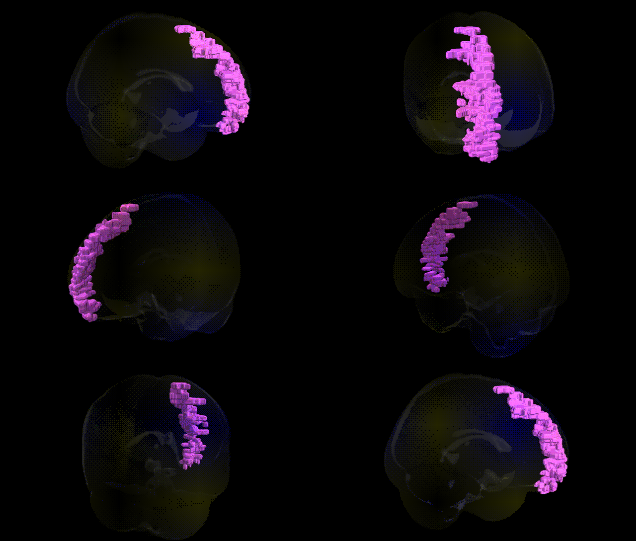
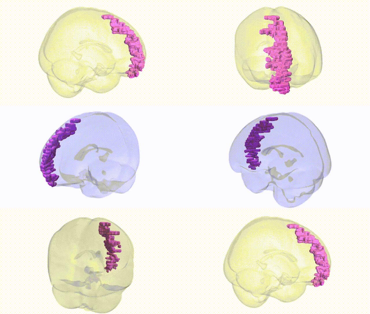
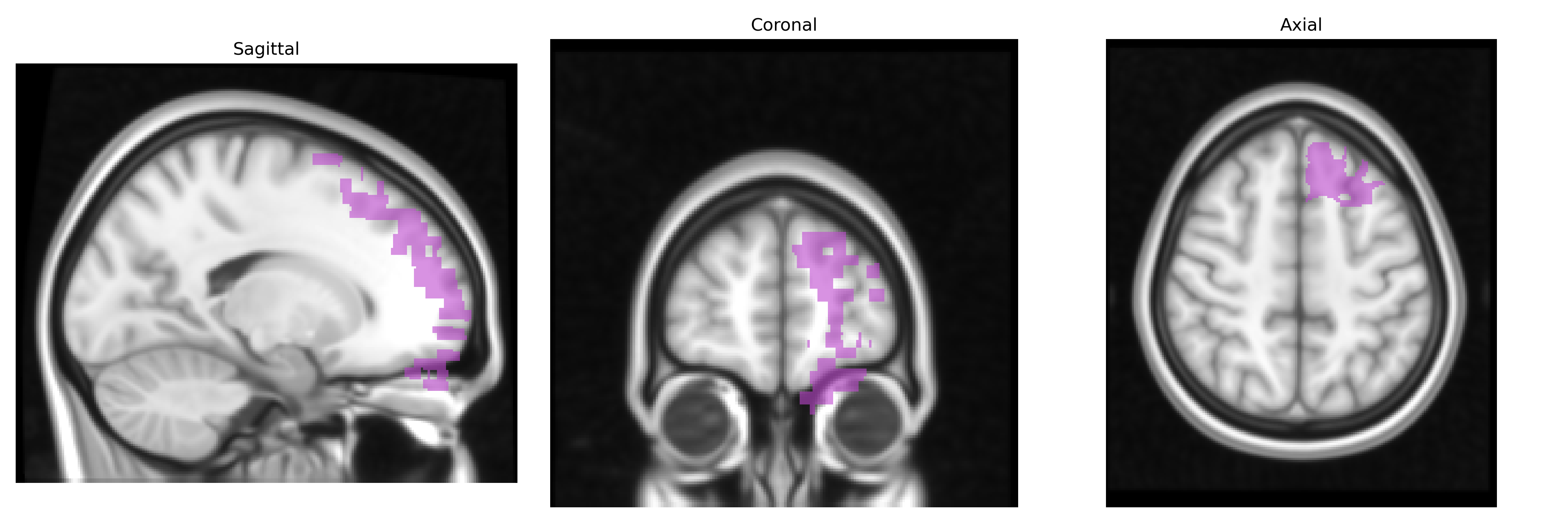
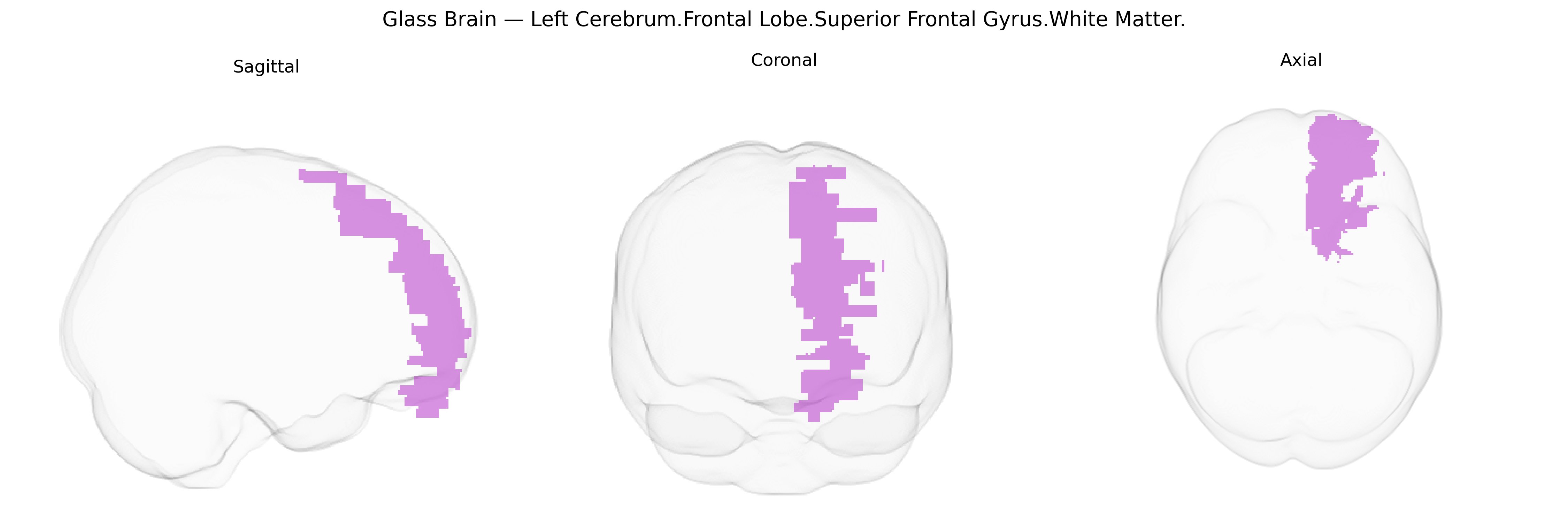
The bilateral Left Cerebrum.Frontal Lobe.Superior Frontal Gyrus.White Matter corresponds to the subcortical myelinated fiber pathways underlying the superior frontal gyrus in the dorsomedial frontal lobe. This white matter region carries long association fibers connecting prefrontal areas with other frontal and parietal regions, as well as commissural and projection fibers linking superior frontal cortex to homologous contralateral regions and subcortical structures, including thalamus and basal ganglia. Functionally, it supports higher-order executive processes, working memory, attention, motor planning, and aspects of self-initiated behavior and cognitive control attributed to the superior frontal cortex and adjacent medial frontal areas. There is no direct Wikipedia article for this exact white matter label; a related cortical structure is the Superior frontal gyrus.
The bilateral Left Cerebrum.Frontal Lobe.Superior Frontal Gyrus.White Matter, as defined in the Talairach 1 mm atlas, corresponds largely to dorsal medial and superior prefrontal white matter pathways that have been repeatedly implicated in imaging‑genetics and GWAS studies of brain structure, cognition, and psychiatric risk. Polygenic influences from genes involved in axon guidance, myelination, and synaptic plasticity—notably variants within or near genes such as NRG1, ERBB4, CNTNAP2, DISC1, BDNF, and multiple oligodendrocyte-related loci—have been associated with altered frontal white matter integrity (e.g., fractional anisotropy) in diffusion MRI, particularly in superior frontal and adjacent callosal tracts. Large-scale ENIGMA and UK Biobank GWAS of cortical surface area, thickness, and white matter microstructure identify numerous loci (including MAPT, genes in the major histocompatibility complex, and multiple neuronal development genes) that show effects in or near superior frontal regions, linking these genetic variants to traits such as general cognitive ability, educational attainment, processing speed, and executive function. Psychiatric and neurodevelopmental GWAS—especially for schizophrenia, bipolar disorder, major depressive disorder, ADHD, and autism—have demonstrated that their polygenic risk scores correlate with reduced integrity or altered development of superior frontal and medial prefrontal white matter, suggesting that genetic architectures conferring risk for these disorders partially act via disruption of long-range frontally anchored association fibers. In parallel, genes influencing vascular and metabolic risk (e.g., APOE and other lipid/metabolism loci) have been linked to frontal white matter changes and lesion burden, tying the region’s genetic architecture to susceptibility for small-vessel disease, late-life cognitive decline, and dementia.
Overview generated by GPT-4o (2026).
Region ID: 155
Hemisphere: bilateral
Atlas: Talairach labels 1mm
Full Quality Version: Download MP4
Full Quality Version: Download MP4
Wali Sidiqyar*, Gaurav Rudravaram*, Elyssa M. McMaster, Trent M. Schwartz, Adam M. Saunders, Kurt G. Schilling, Bennett A. Landman "Introducing SPINS: A Shared Public Visualization Library of Neuroanatomical Structures." Medical Imaging with Deep Learning- short paper
This resource is licensed under CC0 1.0 Universal (Public Domain).