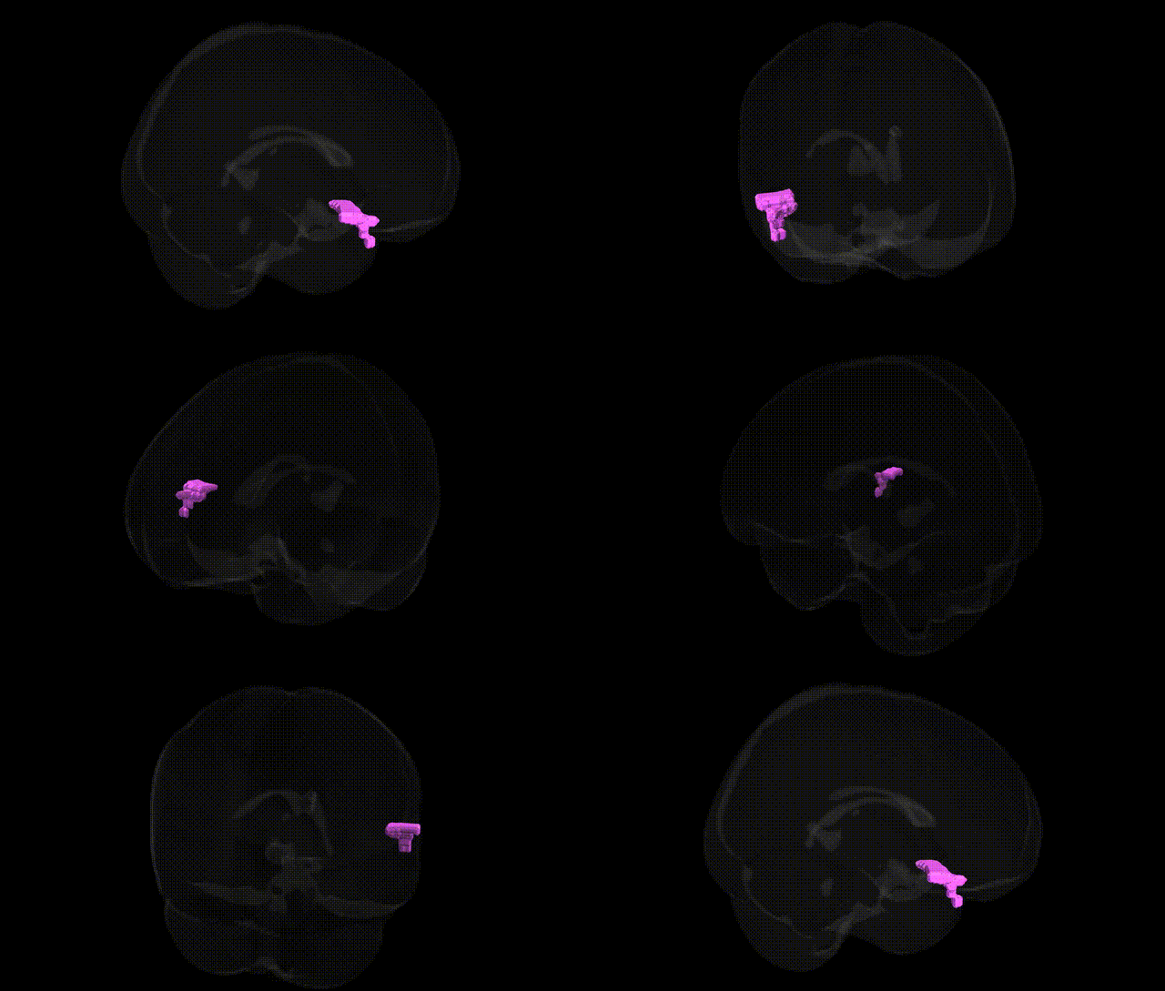
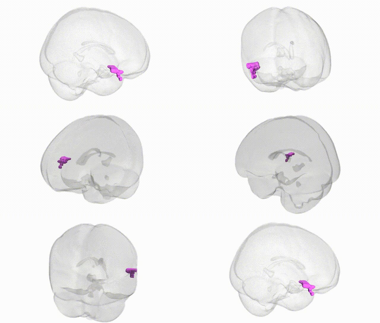
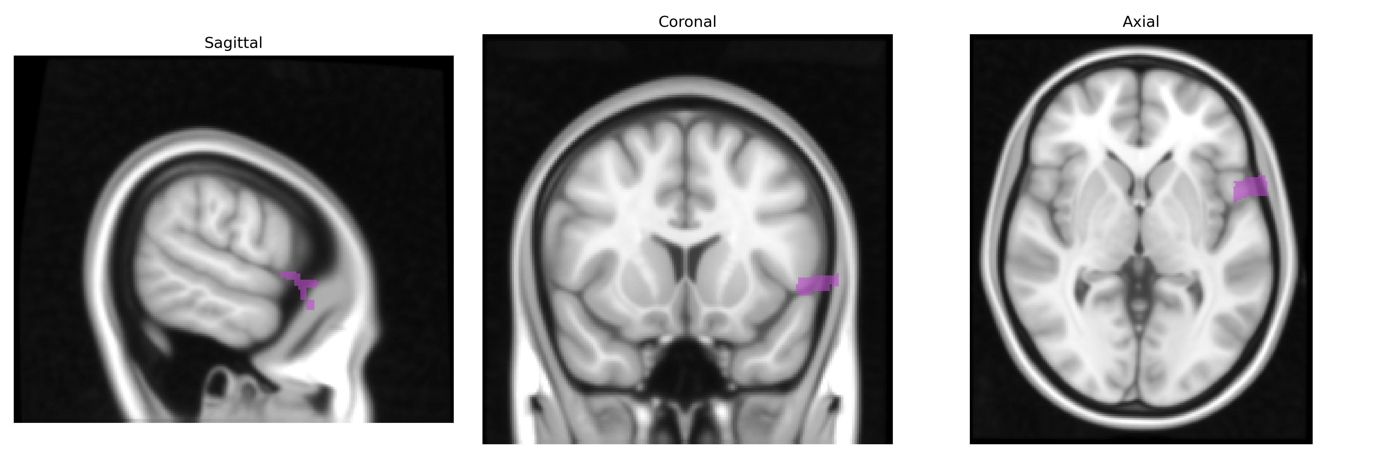
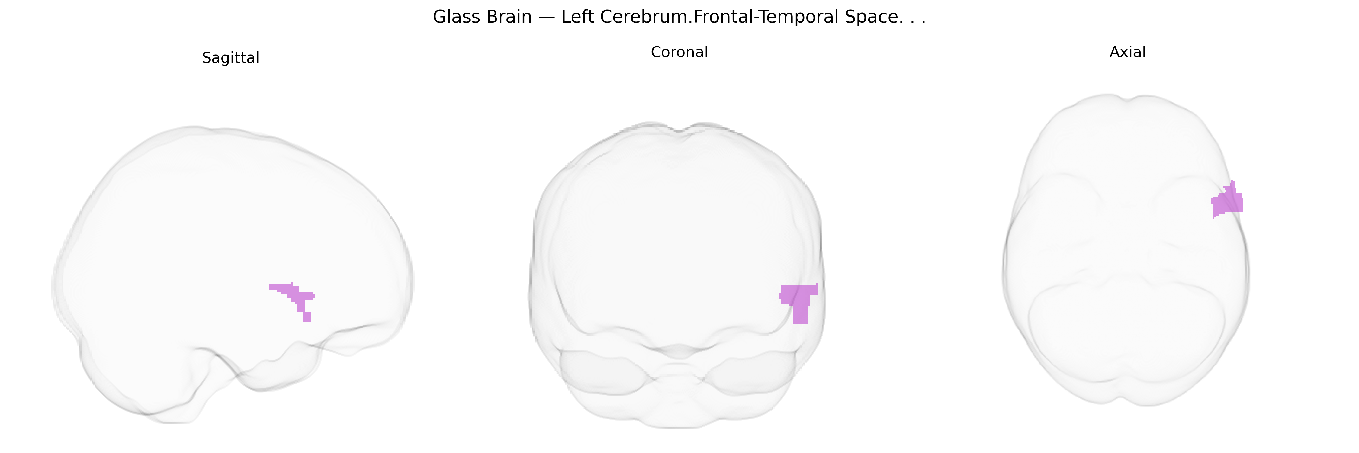
The bilateral Left Cerebrum.Frontal-Temporal space, as defined in the Talairach 1 mm Atlas, denotes a composite region spanning portions of the frontal and temporal lobes within the left cerebral hemisphere. This space typically encompasses areas implicated in higher-order cognitive and executive functions (frontal lobe) and language, auditory processing, and aspects of memory (temporal lobe). Anatomically, it lies anterior to the central sulcus and extends inferiorly and posteriorly toward the superior and middle temporal gyri, overlapping distributed cortical territories rather than a single cytoarchitectonic field. Functionally, activity in this region often reflects integration of executive control, working memory, and language-related processes, including speech production and comprehension, as well as multimodal association functions. There is no direct link for this exact composite label; a related structure is the Cerebral cortex.
The bilateral Left Cerebrum.Frontal-Temporal Space, a macroanatomical zone spanning frontal and temporal cortices in the Talairach 1 mm atlas, overlaps several regions for which GWAS and imaging-genetics studies have identified robust genetic associations, particularly involving cortical thickness, surface area, and gyrification. Common variants in genes related to neurodevelopment and synaptic function (e.g., MIR137, GRIN2B, CNTNAP2, DISC1, FOXP2, and CACNA1C) influence structural and functional properties of inferior frontal and superior/middle temporal areas, which together support language, social cognition, and executive control. Large consortia such as ENIGMA and UK Biobank have reported that polygenic scores for schizophrenia, bipolar disorder, and major depression are associated with altered gray-matter volume and cortical thickness in frontotemporal territories, while Alzheimer’s disease risk variants (notably APOE ε4 and CLU) show associations with atrophy patterns extending into lateral temporal and frontal association cortex. Autism spectrum disorder, specific language impairment, and developmental dyslexia GWAS and candidate-gene studies highlight genes affecting frontotemporal circuitry and white-matter connectivity (e.g., DCDC2, KIAA0319, ROBO1), often mapping via imaging to structural or functional differences in perisylvian frontal–temporal networks. Additionally, common variants discovered in brain-structure GWAS (e.g., in HMGA2, IGF1, SLC39A8, and other loci regulating growth and neuronal differentiation) show associations with global and regional frontotemporal morphology, indicating that this bilateral frontal–temporal space is a key target of polygenic influences on both normal neuroanatomical variation and vulnerability to psychiatric and neurodegenerative disorders.
Overview generated by GPT-4o (2026).
Region ID: 394
Hemisphere: bilateral
Atlas: Talairach labels 1mm
Full Quality Version: Download MP4
Full Quality Version: Download MP4
Wali Sidiqyar*, Gaurav Rudravaram*, Elyssa M. McMaster, Trent M. Schwartz, Adam M. Saunders, Kurt G. Schilling, Bennett A. Landman "Introducing SPINS: A Shared Public Visualization Library of Neuroanatomical Structures." Medical Imaging with Deep Learning- short paper
This resource is licensed under CC0 1.0 Universal (Public Domain).