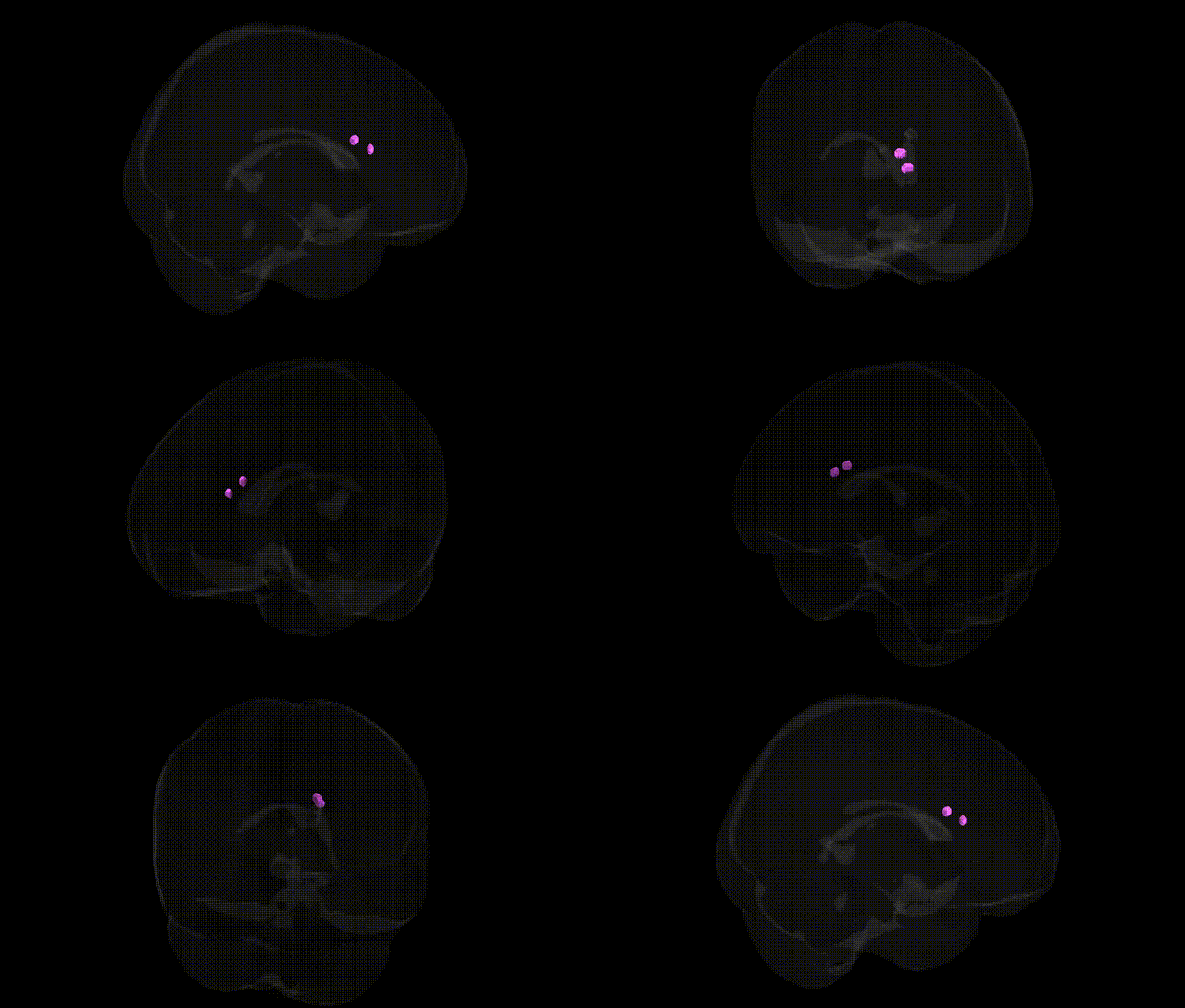
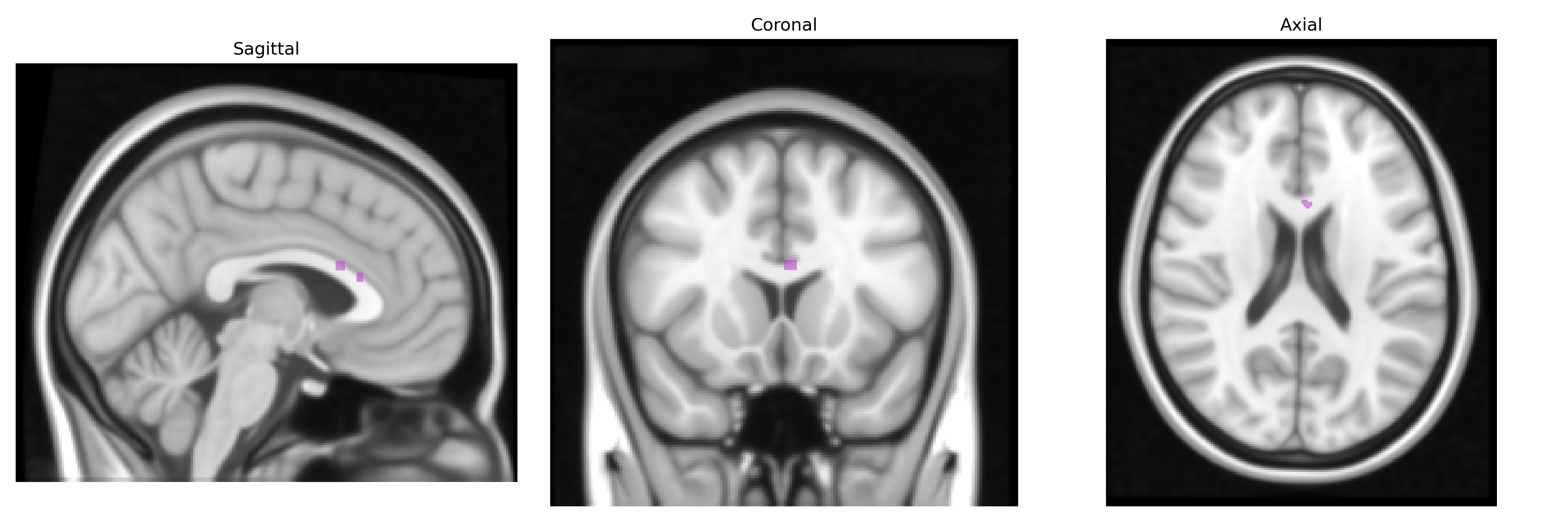
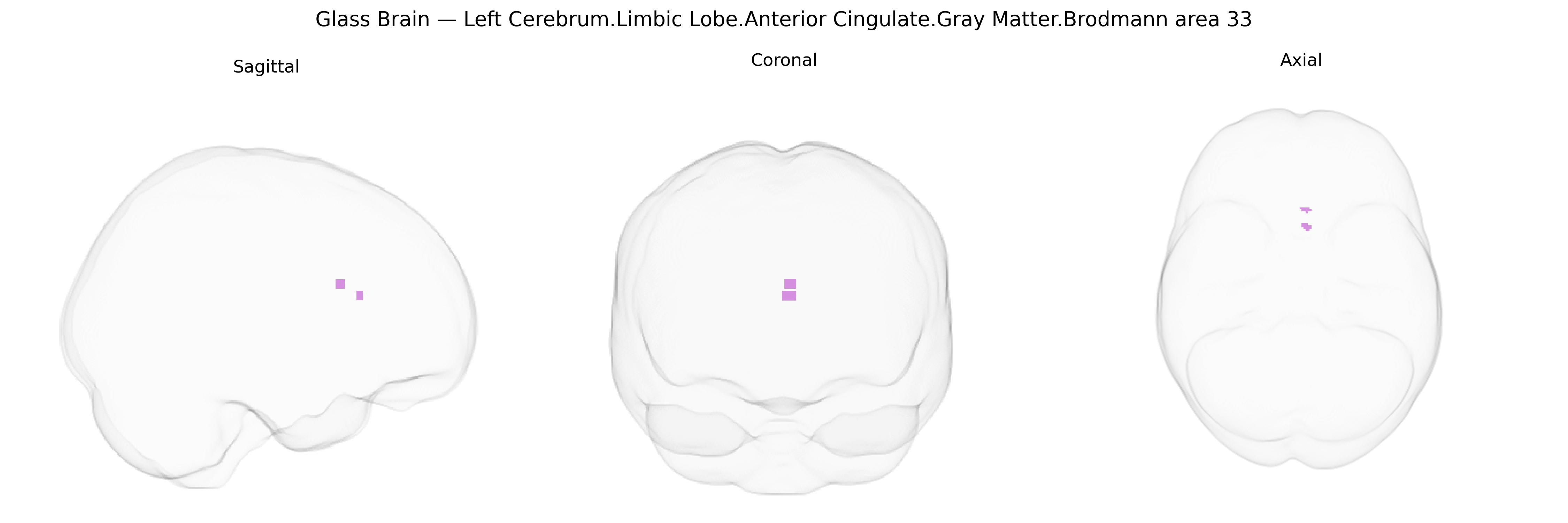
The bilateral Left Cerebrum Limbic Lobe Anterior Cingulate Gray Matter Brodmann area 33 (BA33) is a small cytoarchitectonic subdivision of the anterior cingulate cortex located on the medial surface of the frontal lobe, deep within the cingulate sulcus and adjacent to other anterior cingulate areas such as BA24 and BA32. It is composed of agranular cortex and forms part of the limbic system, integrating affective, autonomic, and cognitive processes. BA33 receives inputs from prefrontal, limbic, and thalamic regions and projects to other cingulate and limbic structures, contributing to functions such as emotional evaluation, regulation of autonomic responses, and modulation of pain and visceromotor activity. In neuroimaging and stereotactic frameworks like the Talairach atlas, BA33 serves as a reference region for studies of emotion, motivation, and affective components of pain processing.
The bilateral left anterior cingulate cortex (ACC), including Brodmann area 33 within the limbic lobe, has been repeatedly implicated in genetic studies of psychiatric and cognitive traits, although few GWAS target BA33 specifically. Large-scale GWAS of major depressive disorder, schizophrenia, bipolar disorder, and anxiety-related traits often identify risk loci whose imaging-genetic follow-ups show effects on ACC volume, cortical thickness, or functional activation, including variants in genes such as CACNA1C, ZNF804A, DRD2, SLC6A4, and BDNF. For example, serotonin transporter (SLC6A4) polymorphisms and the BDNF Val66Met variant have been associated with altered ACC activation during emotional and pain processing, while schizophrenia- and bipolar-associated loci (e.g., CACNA1C, GRM3) have been linked to ACC structural and connectivity changes in imaging GWAS and candidate-gene studies. ACC gray matter measures, including thickness and surface area derived from MRI, have significant heritability and appear in GWAS of brain morphology, with loci in genes involved in neurodevelopment and synaptic function (e.g., PPP1R17, TCF4, and other cortical-development genes) influencing this region’s structure. Genetic influences on ACC function and morphology have been reported in relation to traits such as neuroticism, harm avoidance, cognitive control, and pain sensitivity, and ACC abnormalities linked to these variants are seen across disorders including depression, PTSD, obsessive-compulsive disorder, and substance use, suggesting that risk alleles in neurodevelopmental and neurotransmission pathways exert pleiotropic effects that converge on anterior cingulate circuitry encompassing BA33.
Overview generated by GPT-4o (2026).
Region ID: 838
Hemisphere: bilateral
Atlas: Talairach labels 1mm
Full Quality Version: Download MP4
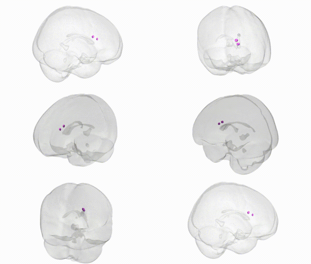
Full Quality Version: Download MP4
Wali Sidiqyar*, Gaurav Rudravaram*, Elyssa M. McMaster, Trent M. Schwartz, Adam M. Saunders, Kurt G. Schilling, Bennett A. Landman "Introducing SPINS: A Shared Public Visualization Library of Neuroanatomical Structures." Medical Imaging with Deep Learning- short paper
This resource is licensed under CC0 1.0 Universal (Public Domain).