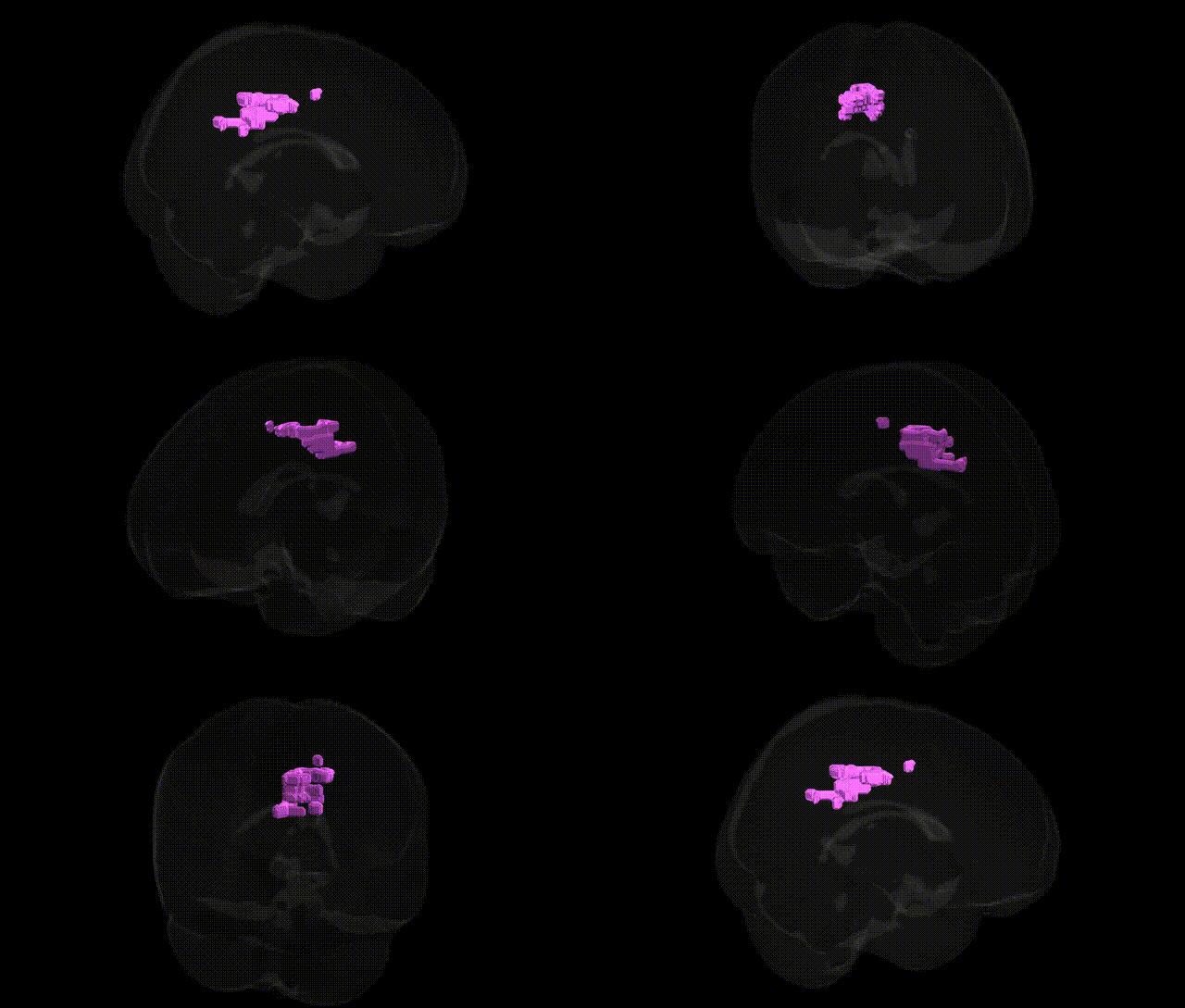
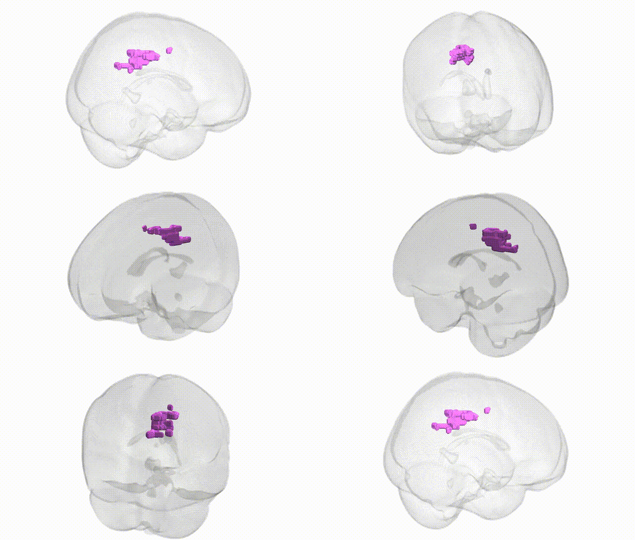
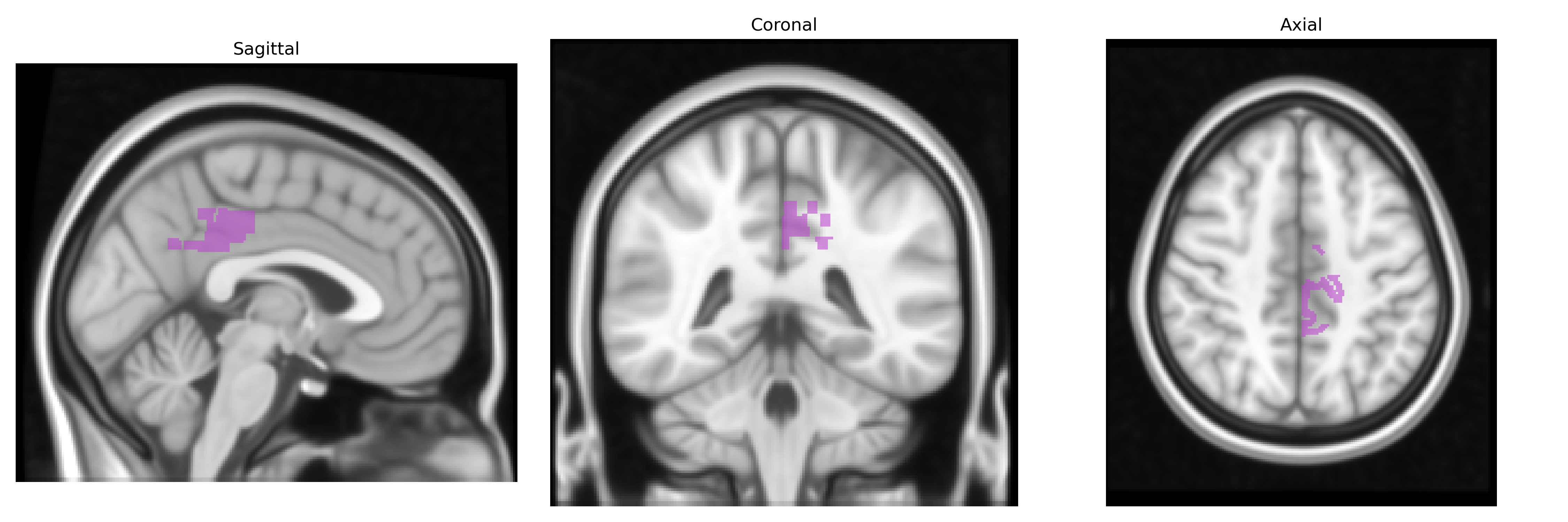
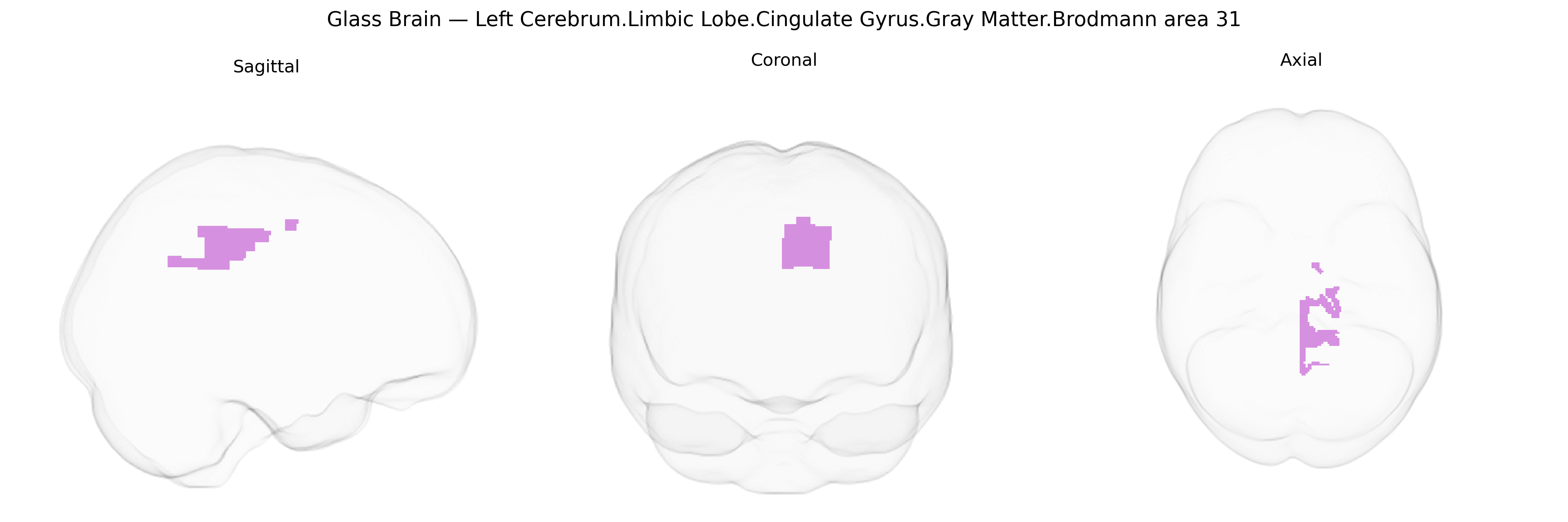
Bilateral Left Cerebrum.Limbic Lobe.Cingulate Gyrus.Gray Matter.Brodmann area 31 corresponds to a posterior segment of the cingulate gyrus situated in the medial parietal cortex along the banks of the cingulate sulcus, extending into the posterior cingulate and precuneus regions. Cytoarchitectonically, it is an association cortex area characterized by relatively prominent granular layers and dense corticocortical connections, integrating multimodal sensory, mnemonic, and attentional information. Functionally, Brodmann area 31 is implicated in episodic memory retrieval, self-referential and internally directed thought, visuospatial processing, and aspects of emotional evaluation, and forms part of large-scale networks such as the default mode network. Lesions or altered activity in this region have been associated with disturbances in memory, altered consciousness, and affective or attentional dysfunctions. Brodmann area 31
Brodmann area 31 in the posterior cingulate/medial parietal cortex has been repeatedly implicated in genetic studies of neuropsychiatric and neurodegenerative traits, although most GWAS map loci to broader posterior cingulate or default mode network regions rather than Talairach-specific BA31. Twin and heritability studies show substantial genetic influence on cingulate gray matter volume and thickness, with SNP-based heritability estimates often in the 20–40% range. Large neuroimaging–genetics consortia (e.g., ENIGMA, UK Biobank) have identified common variants near genes involved in synaptic function, neurodevelopment, and myelination that associate with cortical thickness and surface area in posterior cingulate/precuneus regions encompassing BA31. GWAS and imaging–genetics work in Alzheimer’s disease and mild cognitive impairment link APOE and other late-onset AD risk loci to atrophy and hypometabolism in posterior cingulate/BA31, matching this region’s role as an early site of amyloid and tau-related degeneration. Schizophrenia and major depressive disorder polygenic risk scores correlate with structural and functional alterations in the posterior cingulate, while specific variants (e.g., in CACNA1C, ZNF804A, and other synaptic/neurodevelopmental genes) have been reported to influence cingulate connectivity or activation in case–control and intermediate-phenotype studies. Additional associations connect posterior cingulate/BA31 structure and function to genetic risk for autism spectrum traits, attention-deficit/hyperactivity disorder, and personality dimensions such as neuroticism and harm avoidance, as well as to general cognitive performance and memory-related traits, highlighting this region as a genetically modulated hub for default mode network activity, self-referential processing, and memory.
Overview generated by GPT-4o (2026).
Region ID: 915
Hemisphere: bilateral
Atlas: Talairach labels 1mm
Full Quality Version: Download MP4
Full Quality Version: Download MP4
Wali Sidiqyar*, Gaurav Rudravaram*, Elyssa M. McMaster, Trent M. Schwartz, Adam M. Saunders, Kurt G. Schilling, Bennett A. Landman "Introducing SPINS: A Shared Public Visualization Library of Neuroanatomical Structures." Medical Imaging with Deep Learning- short paper
This resource is licensed under CC0 1.0 Universal (Public Domain).