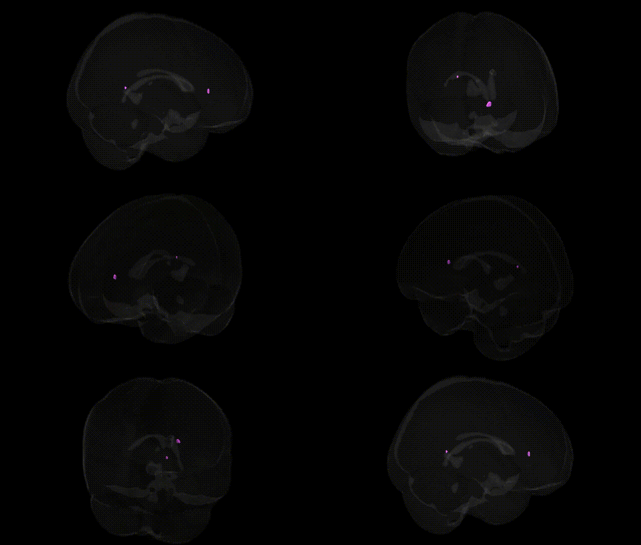
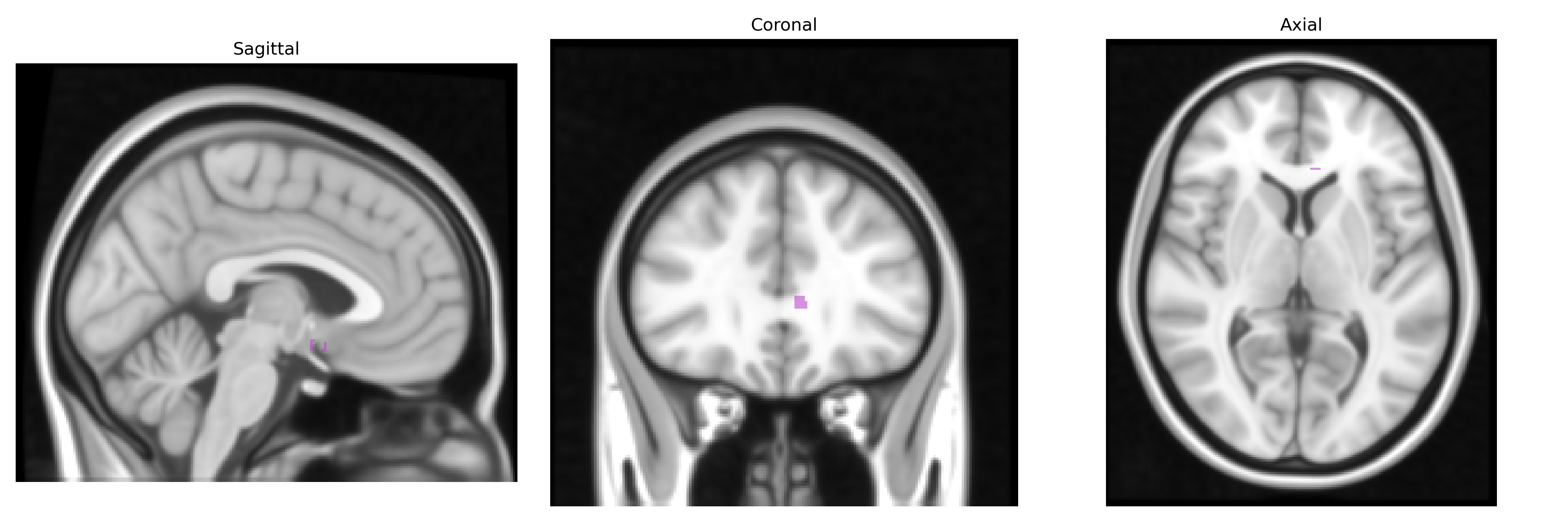
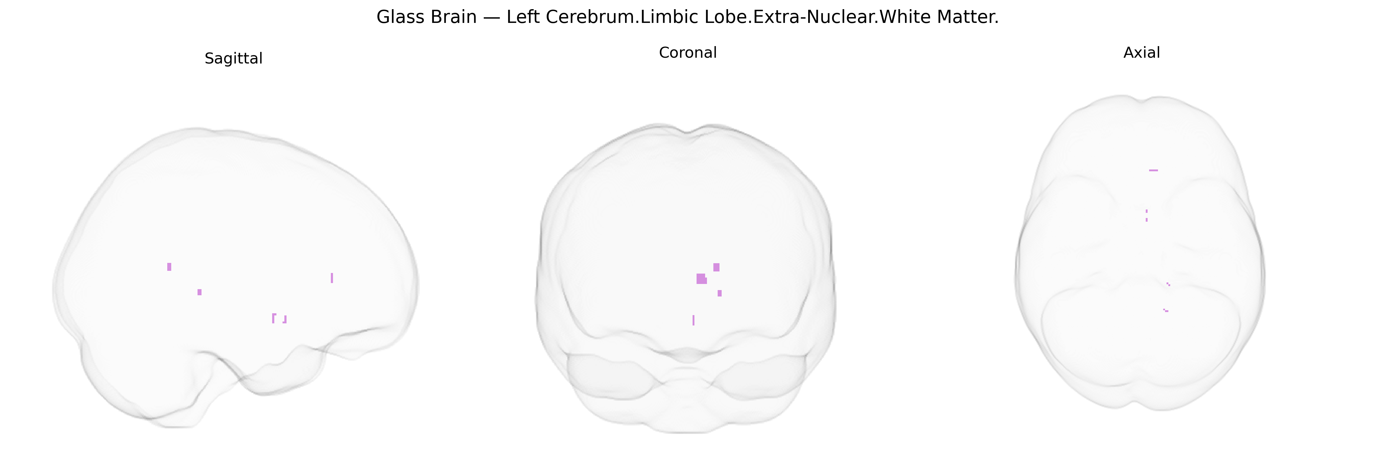
The bilateral Left Cerebrum.Limbic Lobe.Extra-Nuclear.White Matter region in the Talairach 1 mm atlas refers to limbic-associated white matter tracts located outside the primary nuclear (gray matter) structures of the limbic system, such as the hippocampus and amygdala. This designation typically encompasses portions of major fiber pathways—including segments of the cingulum bundle and related association fibers—that interconnect limbic cortices with subcortical limbic nuclei and other cerebral regions, supporting functions such as emotion, memory, and autonomic regulation. As “Extra-Nuclear White Matter” is not a standard anatomical term with its own article, a closely related and representative structure is the cingulum bundle: Cingulum.
The bilateral Left Cerebrum Limbic Lobe Extra-Nuclear White Matter, as defined in the Talairach 1 mm atlas, corresponds broadly to limbic-associated periventricular and deep white matter tracts connecting structures such as the amygdala, hippocampus, basal forebrain, and cingulate regions, and genetic associations are typically inferred from studies of limbic and periventricular white matter rather than this specific atlas label. Large GWAS of white matter microstructure (e.g., using diffusion MRI measures such as fractional anisotropy and mean diffusivity) have identified robust associations with genes implicated in axon guidance, myelination, and oligodendrocyte function, including variants in genes such as MAG, MOG, PLP1, and loci near NRXN1, as well as glutamatergic and synaptic genes; many of these effects are strongest in limbic and frontal white matter tracts. Polygenic influences on this region’s structure and integrity have also been inferred from overlap with risk loci for schizophrenia, bipolar disorder, major depressive disorder, and autism spectrum disorder, in which limbic white matter alterations (especially in tracts near the amygdala–hippocampal complex and cingulum) are common imaging findings. GWAS of white matter hyperintensities, which frequently involve periventricular and limbic-adjacent white matter, have repeatedly implicated loci such as those near COL4A2, PDCD11, PLEKHG1, HTRA1, and genes related to small-vessel disease and vascular integrity, linking this region to genetic risk for stroke, cognitive decline, and late-life dementia. Additional associations involve polygenic scores for educational attainment and general cognitive ability, which correlate with higher integrity of limbic and association white matter, as well as APOE and other Alzheimer’s disease–related loci that influence limbic white matter degeneration. Overall, while no major GWAS has targeted the Talairach-defined “Extra-Nuclear” limbic white matter label specifically, convergent genetic evidence from imaging-genetics and disease GWAS indicates that common variants affecting myelination, vascular health, and neurodevelopmental synaptic pathways contribute to interindividual differences and disease vulnerability in the limbic white matter systems encompassed by this region.
Overview generated by GPT-4o (2026).
Region ID: 370
Hemisphere: bilateral
Atlas: Talairach labels 1mm
Full Quality Version: Download MP4
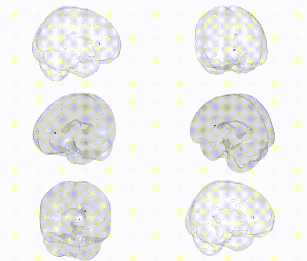
Full Quality Version: Download MP4
Wali Sidiqyar*, Gaurav Rudravaram*, Elyssa M. McMaster, Trent M. Schwartz, Adam M. Saunders, Kurt G. Schilling, Bennett A. Landman "Introducing SPINS: A Shared Public Visualization Library of Neuroanatomical Structures." Medical Imaging with Deep Learning- short paper
This resource is licensed under CC0 1.0 Universal (Public Domain).