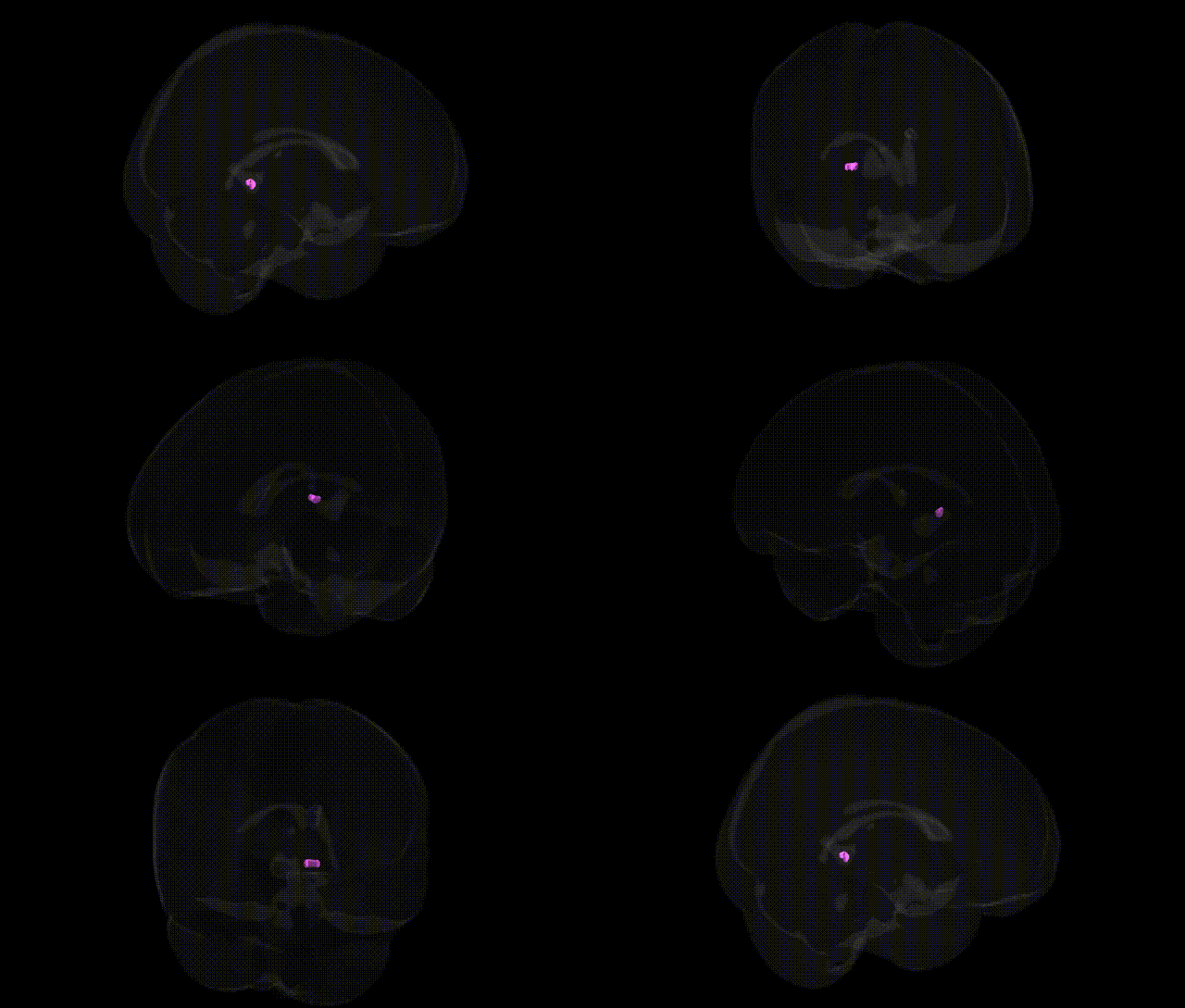
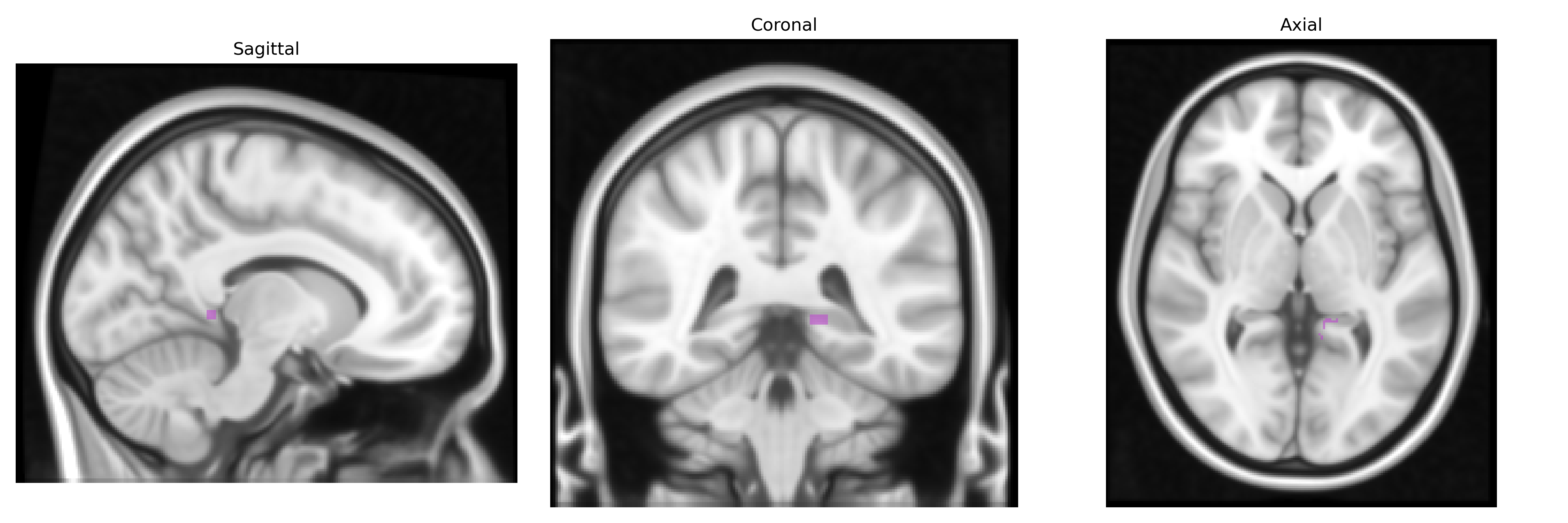
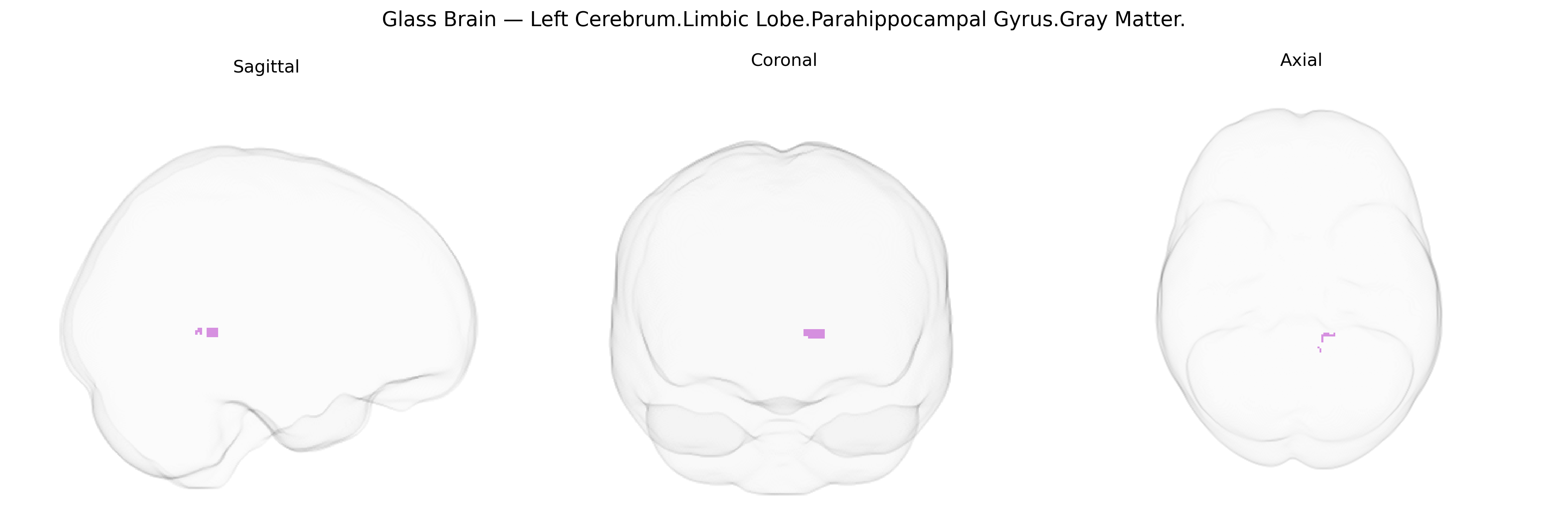
The bilateral Left Cerebrum Limbic Lobe Parahippocampal Gyrus Gray Matter corresponds to cortical gray matter along the medial temporal lobe that forms part of the limbic system, arching around the hippocampal formation. It is primarily involved in contextual and spatial aspects of memory, visuospatial processing, and scene recognition, and contributes to encoding and retrieval of episodic memories as well as navigation within environments. This region receives multimodal input from association cortices and projects to hippocampal and entorhinal regions, integrating sensory and mnemonic information. Its gray matter architecture includes specialized cytoarchitecture adapted for complex associative processing, and it is frequently implicated in temporal lobe epilepsy and neurodegenerative disorders affecting memory networks. Parahippocampal gyrus
The bilateral left parahippocampal gyrus (limbic lobe gray matter) has been repeatedly implicated in genetic studies of memory, emotion, and neuropsychiatric disease, with GWAS and imaging-genetics work highlighting polygenic influences rather than single major loci; variants in genes involved in synaptic plasticity, glutamatergic signaling, and neurodevelopment (such as BDNF, APOE, GRIN2B, and DISC1) have been associated with structural and functional variation in this region, particularly its volume and activation during episodic and spatial memory tasks. Large-scale imaging GWAS (e.g., ENIGMA and UK Biobank) indicate that parahippocampal thickness and gray-matter volume are heritable and share genetic correlations with Alzheimer’s disease, major depressive disorder, and schizophrenia, where risk loci in genes related to immune function (e.g., MHC region), lipid metabolism, and synaptic organization contribute to case–control differences in parahippocampal structure. In Alzheimer’s disease, APOE ε4 and other late-onset AD loci are associated with accelerated atrophy of the medial temporal lobe, including the parahippocampal gyrus, paralleling early episodic memory decline, while in schizophrenia and bipolar disorder, common risk variants aggregate to predict reduced parahippocampal volume and altered connectivity with the hippocampus and prefrontal cortex. Additional GWAS on anxiety traits, post-traumatic stress symptoms, and neuroticism show that polygenic risk scores for these phenotypes correlate with parahippocampal gray-matter differences and limbic network alterations, suggesting that shared genetic architecture influences both structural variation in this region and liability to mood and stress-related disorders.
Overview generated by GPT-4o (2026).
Region ID: 570
Hemisphere: bilateral
Atlas: Talairach labels 1mm
Full Quality Version: Download MP4
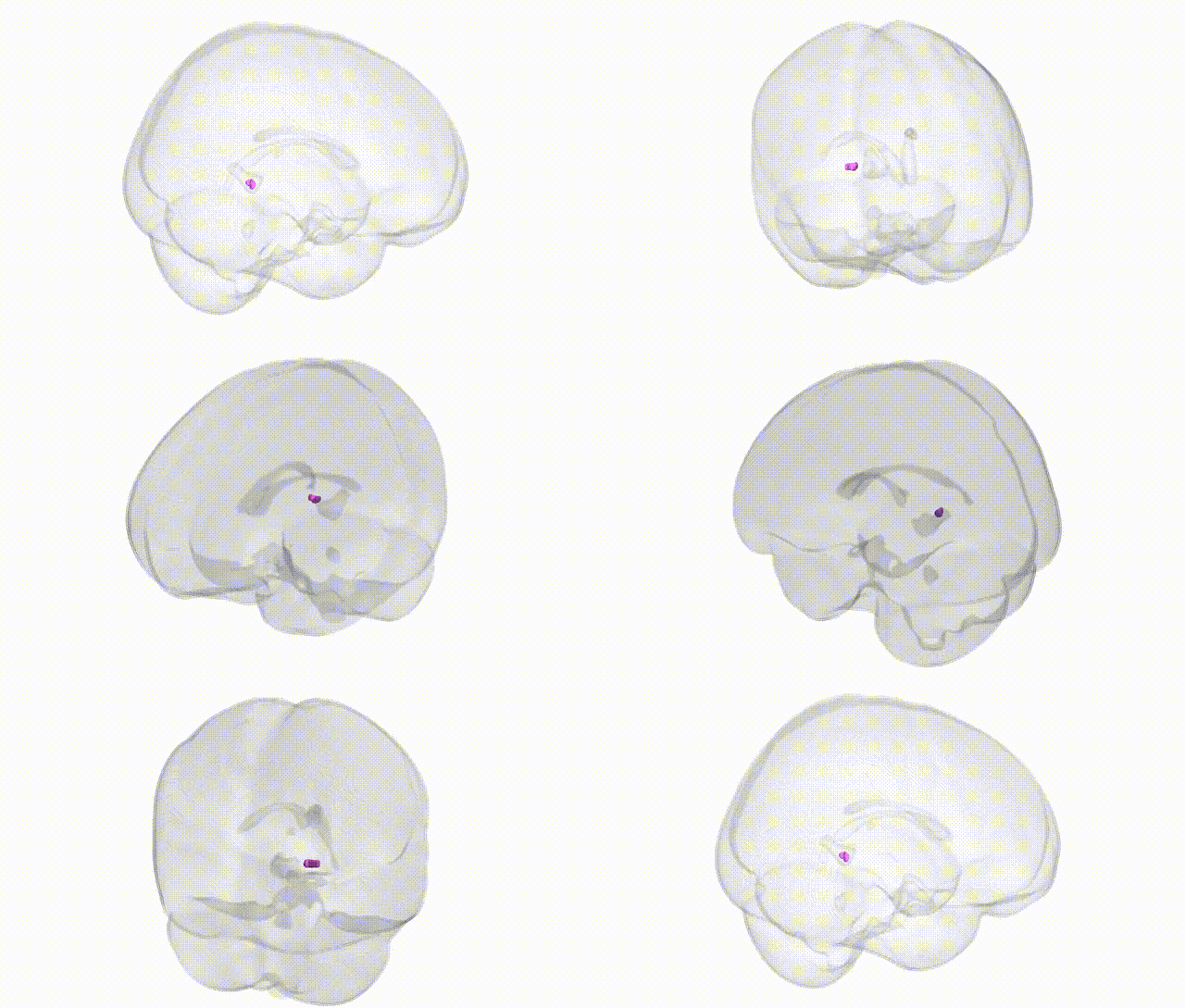
Full Quality Version: Download MP4
Wali Sidiqyar*, Gaurav Rudravaram*, Elyssa M. McMaster, Trent M. Schwartz, Adam M. Saunders, Kurt G. Schilling, Bennett A. Landman "Introducing SPINS: A Shared Public Visualization Library of Neuroanatomical Structures." Medical Imaging with Deep Learning- short paper
This resource is licensed under CC0 1.0 Universal (Public Domain).