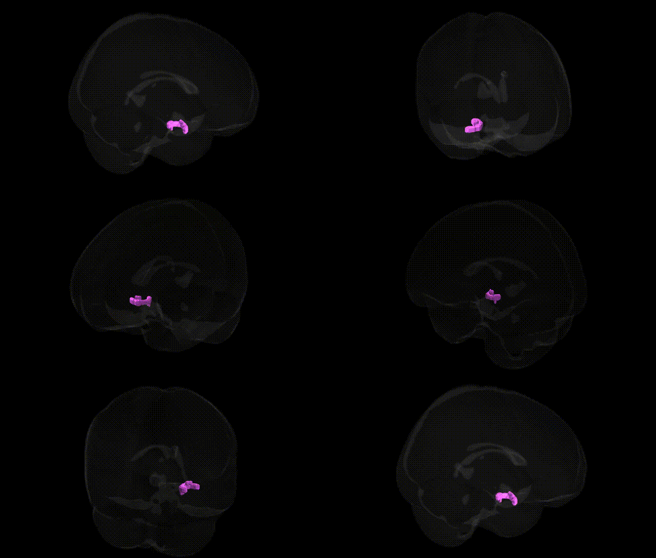
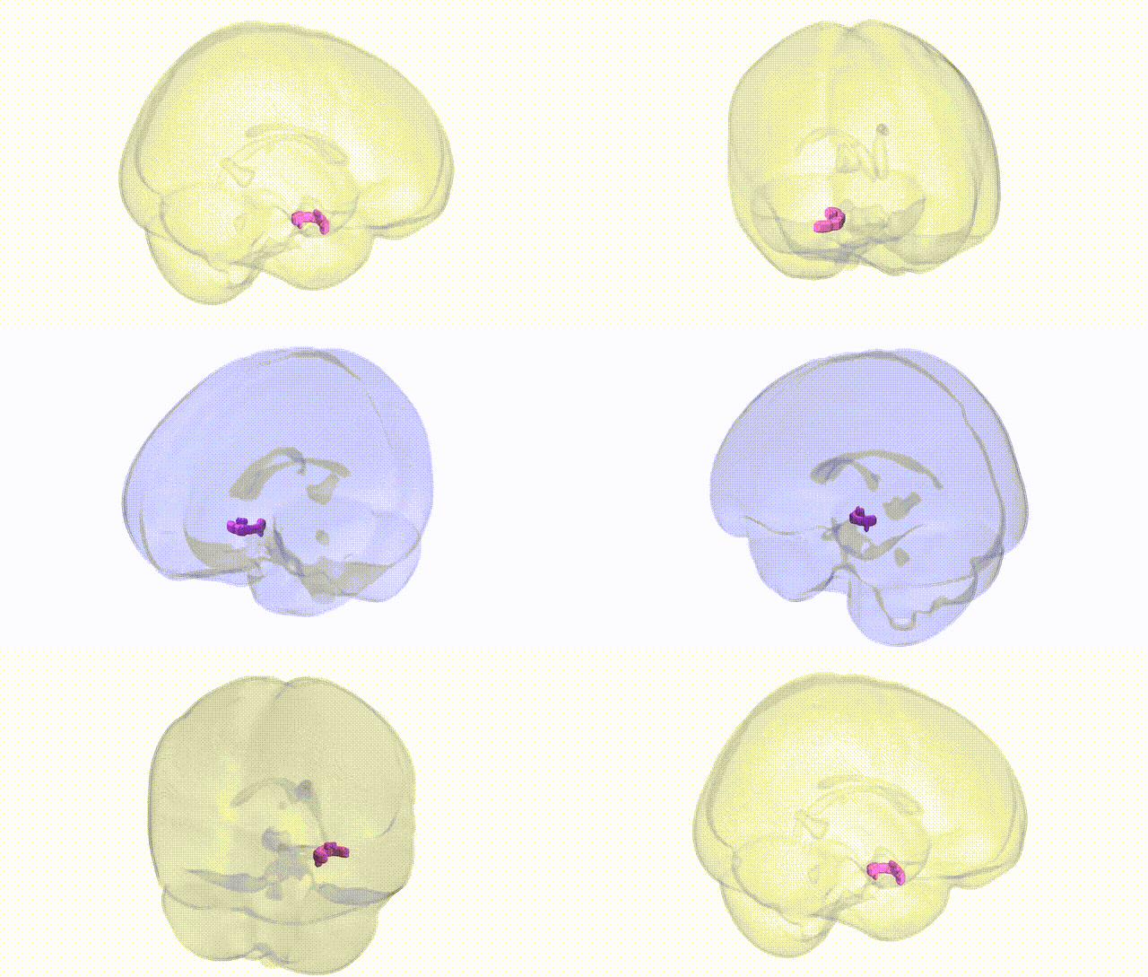
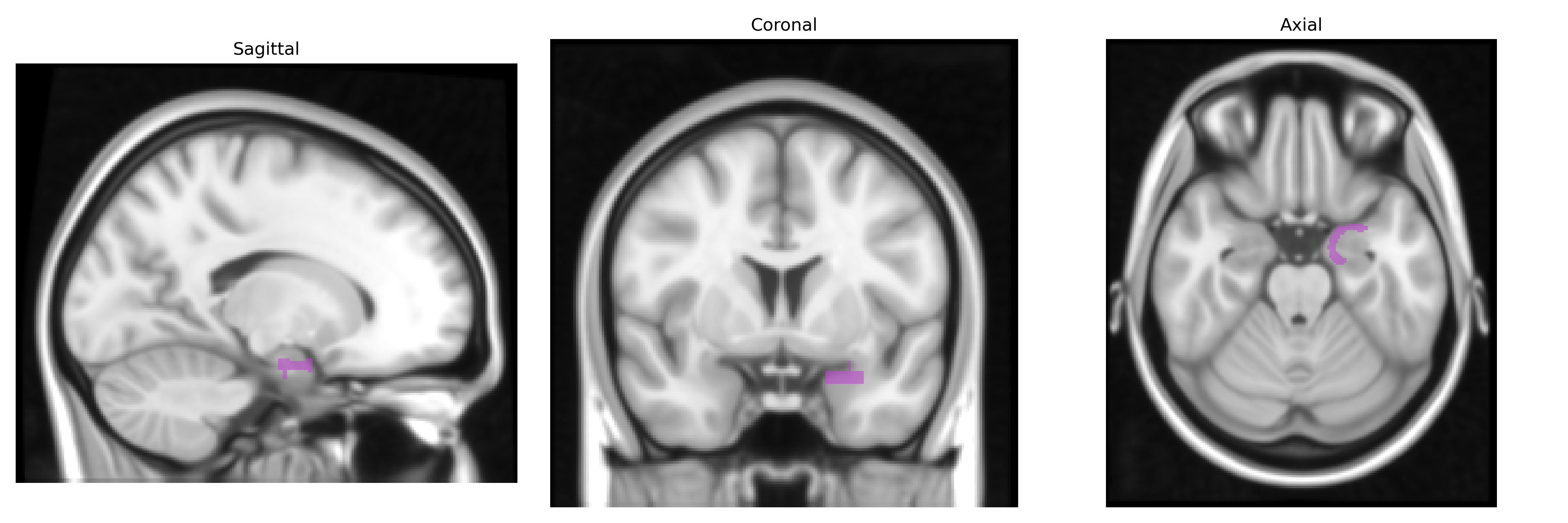
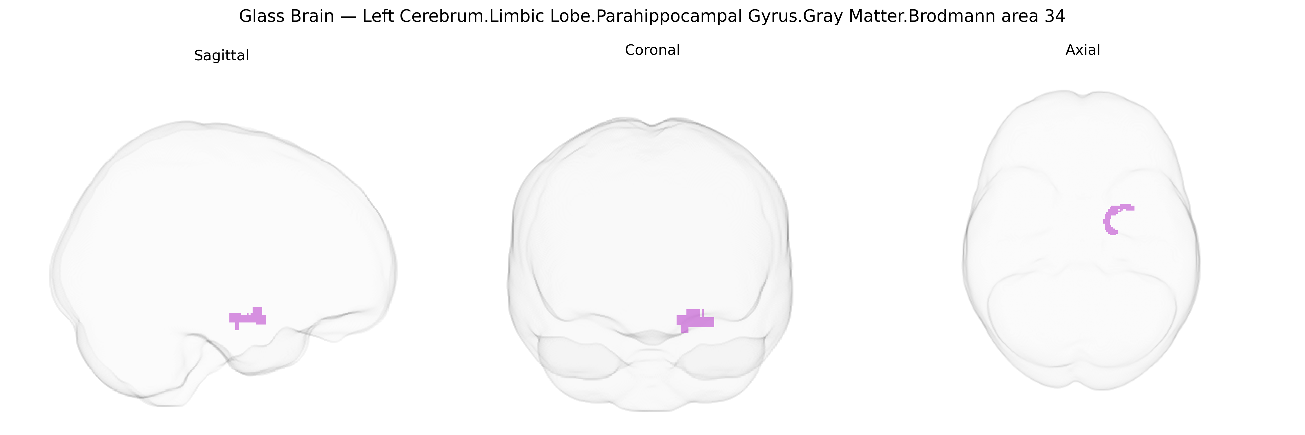
The bilateral Left Cerebrum Limbic Lobe Parahippocampal Gyrus Gray Matter Brodmann area 34 corresponds to a narrow allocortical strip in the anterior medial temporal lobe, commonly identified with the entorhinal cortex, forming a major gateway between the neocortex and hippocampal formation. Located along the anterior parahippocampal gyrus adjacent to the hippocampal head and amygdala, this region integrates high-order multimodal sensory inputs and relays them into hippocampal circuits critical for declarative memory encoding, spatial navigation, and contextual association. Histologically, Brodmann area 34 is characterized by a transitional cortical architecture between three-layered archicortex and six-layered neocortex, with dense layer II islands giving rise to perforant path projections. Functionally, it participates in forming and updating episodic memory representations and is notably vulnerable in early Alzheimer’s disease and other temporal lobe pathologies. There is no direct link for “Brodmann area 34”; a closely related structure is the Entorhinal cortex.
The bilateral parahippocampal gyrus gray matter in Brodmann area 34, as defined in the Talairach 1 mm atlas, has been implicated in multiple genetic and GWAS findings largely through imaging–genetics and neuropsychiatric studies that map variants to medial temporal lobe structure and function. Common polymorphisms in genes related to synaptic plasticity, neurodevelopment, and neurodegeneration—such as APOE (particularly ε4), BDNF (e.g., Val66Met), and CLU, CR1, and other Alzheimer’s disease risk loci—have been associated with parahippocampal and adjacent entorhinal/hippocampal volume, cortical thickness, or metabolic activity, often in the context of memory performance and risk for Alzheimer’s disease or mild cognitive impairment. Large-scale imaging GWAS (e.g., ENIGMA and UK Biobank) have identified significant SNP-based heritability for medial temporal structures including parahippocampal regions and have mapped loci near or within genes involved in axon guidance, neuronal signaling, and immune function (such as variants in or near genes like TESC, SLC39A8, and others) to variation in regional gray matter volumes, although most studies do not isolate BA34 specifically but rather parahippocampal or entorhinal ROIs encompassing it. Genetic associations implicating this region have been reported in schizophrenia, major depression, and bipolar disorder through reduced parahippocampal volume or thickness linked to polygenic risk scores and specific risk variants (e.g., in CACNA1C, ZNF804A, or MHC-region loci), as well as in anxiety-related traits and PTSD, where genetic risk for heightened amygdala–hippocampal reactivity often co-occurs with structural or functional differences in medial temporal lobe circuitry that includes BA34. Collectively, evidence indicates that BA34-linked parahippocampal gray matter is a heritable, genetically modulated region central to memory, emotional processing, and vulnerability to neurodegenerative and psychiatric disorders, though fine-grained, BA34-specific GWAS associations remain comparatively sparse and typically inferred from broader parahippocampal or entorhinal measures.
Overview generated by GPT-4o (2026).
Region ID: 177
Hemisphere: bilateral
Atlas: Talairach labels 1mm
Full Quality Version: Download MP4
Full Quality Version: Download MP4
Wali Sidiqyar*, Gaurav Rudravaram*, Elyssa M. McMaster, Trent M. Schwartz, Adam M. Saunders, Kurt G. Schilling, Bennett A. Landman "Introducing SPINS: A Shared Public Visualization Library of Neuroanatomical Structures." Medical Imaging with Deep Learning- short paper
This resource is licensed under CC0 1.0 Universal (Public Domain).