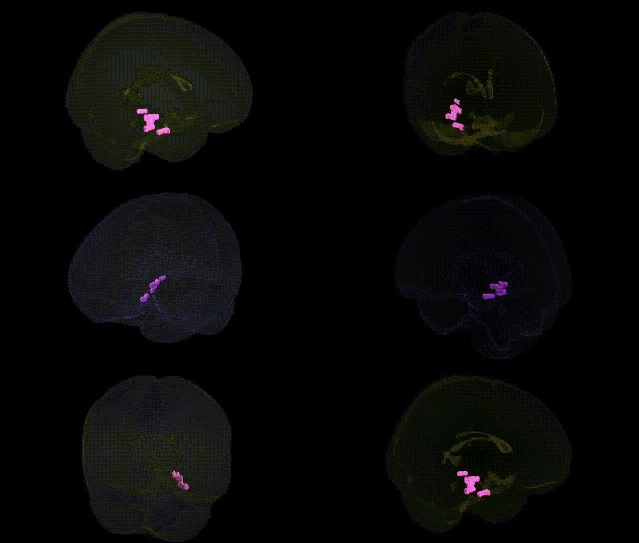
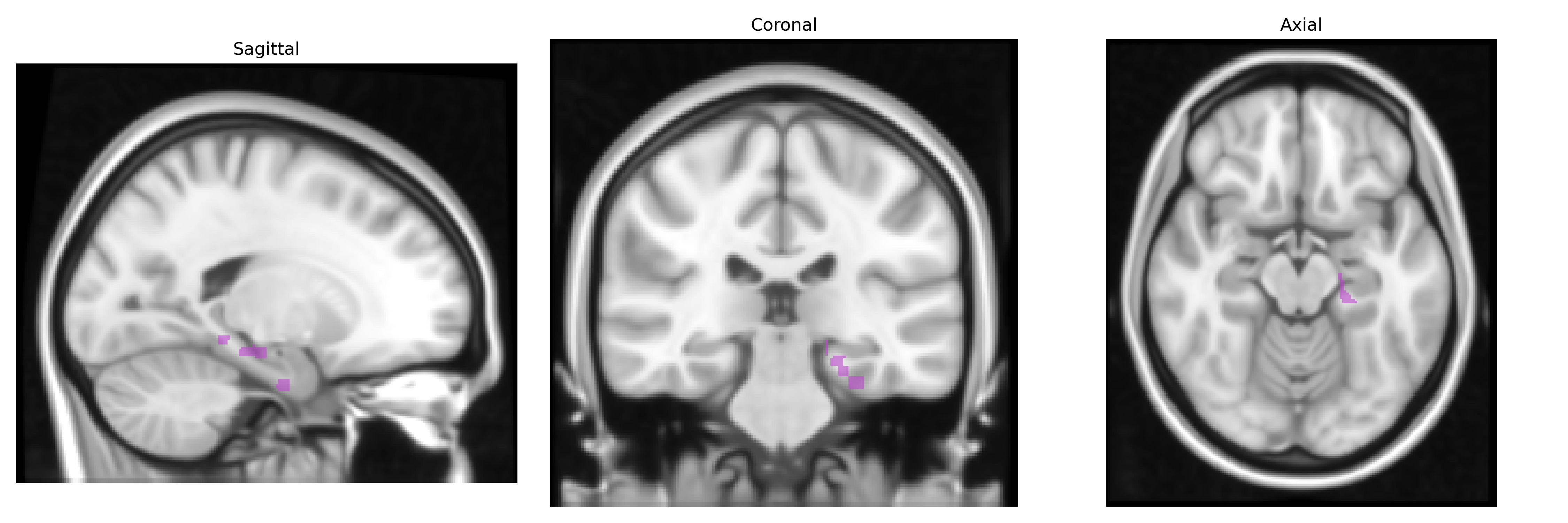
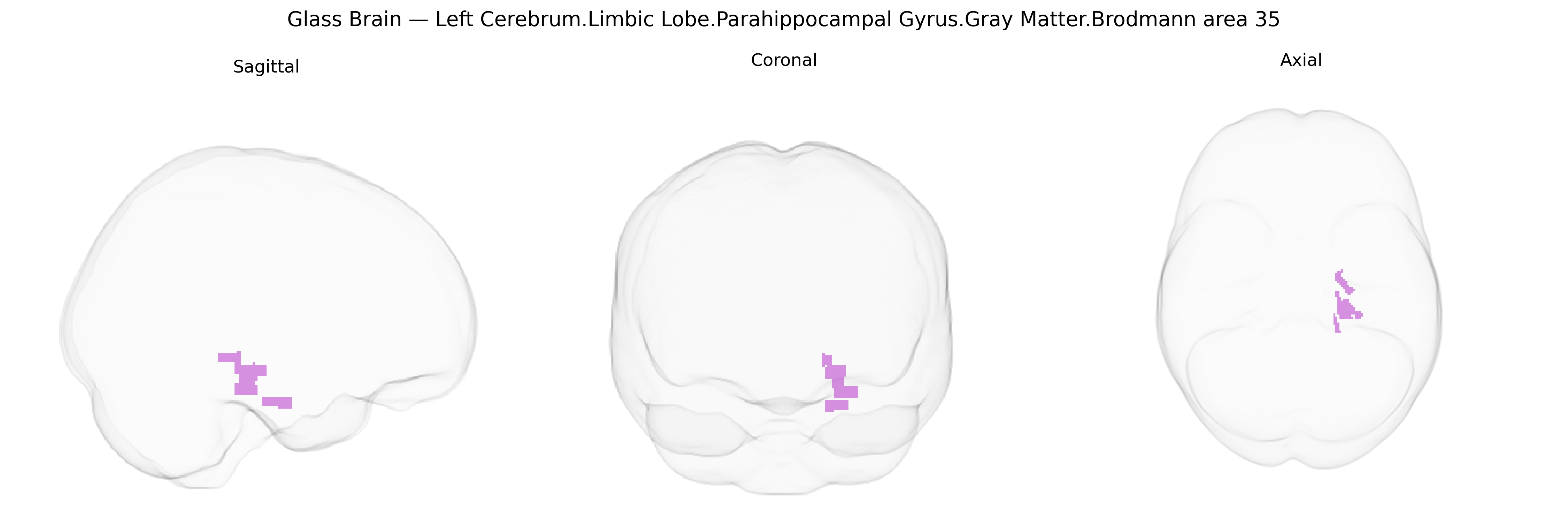
The bilateral Left Cerebrum.Limbic Lobe.Parahippocampal Gyrus.Gray Matter.Brodmann area 35 corresponds to a cytoarchitectonic field located in the anterior–medial portion of the parahippocampal gyrus within the limbic lobe, often considered part of the perirhinal cortex and closely associated with the medial temporal lobe memory system. This region receives polymodal sensory input and is critically involved in recognition memory, particularly familiarity-based processes, associative memory, and the integration of visual object information with contextual and mnemonic cues. It maintains strong reciprocal connections with the hippocampal formation, entorhinal cortex, amygdala, and higher-order visual association cortices, supporting its role in encoding, consolidation, and retrieval of episodic and semantic information. Lesions or dysfunction in Brodmann area 35 have been implicated in memory impairments and are frequently involved in early pathological changes in neurodegenerative conditions such as Alzheimer’s disease due to its proximity to and connectivity with entorhinal and hippocampal regions. There is no direct link for this specific Brodmann area; a closely related structure is the Parahippocampal gyrus.
The bilateral left parahippocampal gyrus gray matter in Brodmann area 35 (often overlapping the perirhinal cortex) has been implicated genetically through its consistent involvement in memory, emotion, and temporal-limbic circuitry, with multiple GWAS and imaging-genetics studies linking variants in Alzheimer’s disease, temporal lobe epilepsy, and mood and anxiety disorders to structure and function in this region. Common Alzheimer’s disease risk genes—such as APOE (particularly ε4), CLU, PICALM, CR1, BIN1, and TREM2—have been associated with parahippocampal and entorhinal/BA35 atrophy, altered cortical thickness, and early neurodegenerative changes, while MAPT and GRN variants contribute to medial temporal and limbic atrophy patterns in frontotemporal dementia. Schizophrenia, bipolar disorder, and major depression GWAS loci (including CACNA1C, ZNF804A, and others in synaptic and calcium-channel pathways) have been related to abnormal gray-matter volume or connectivity in the parahippocampal and adjacent medial temporal regions, and epilepsy genetics (e.g., variants in SCN1A, LGI1, and other channel and synaptic genes) have been connected to mesial temporal sclerosis affecting BA35-containing tissue. Large-scale brain-structure GWAS consortia (such as ENIGMA and UK Biobank) report heritable variation in parahippocampal volume influenced by loci near genes involved in neurodevelopment, axon guidance, and synaptic plasticity (e.g., genes related to Wnt signaling and cell-adhesion molecules), indicating that BA35 morphology and vulnerability partly reflect polygenic influences that overlap with risk architectures for neurodegeneration, epilepsy, and psychiatric illness, even though few variants are specific to BA35 as defined by the Talairach 1 mm atlas.
Overview generated by GPT-4o (2026).
Region ID: 140
Hemisphere: bilateral
Atlas: Talairach labels 1mm
Full Quality Version: Download MP4
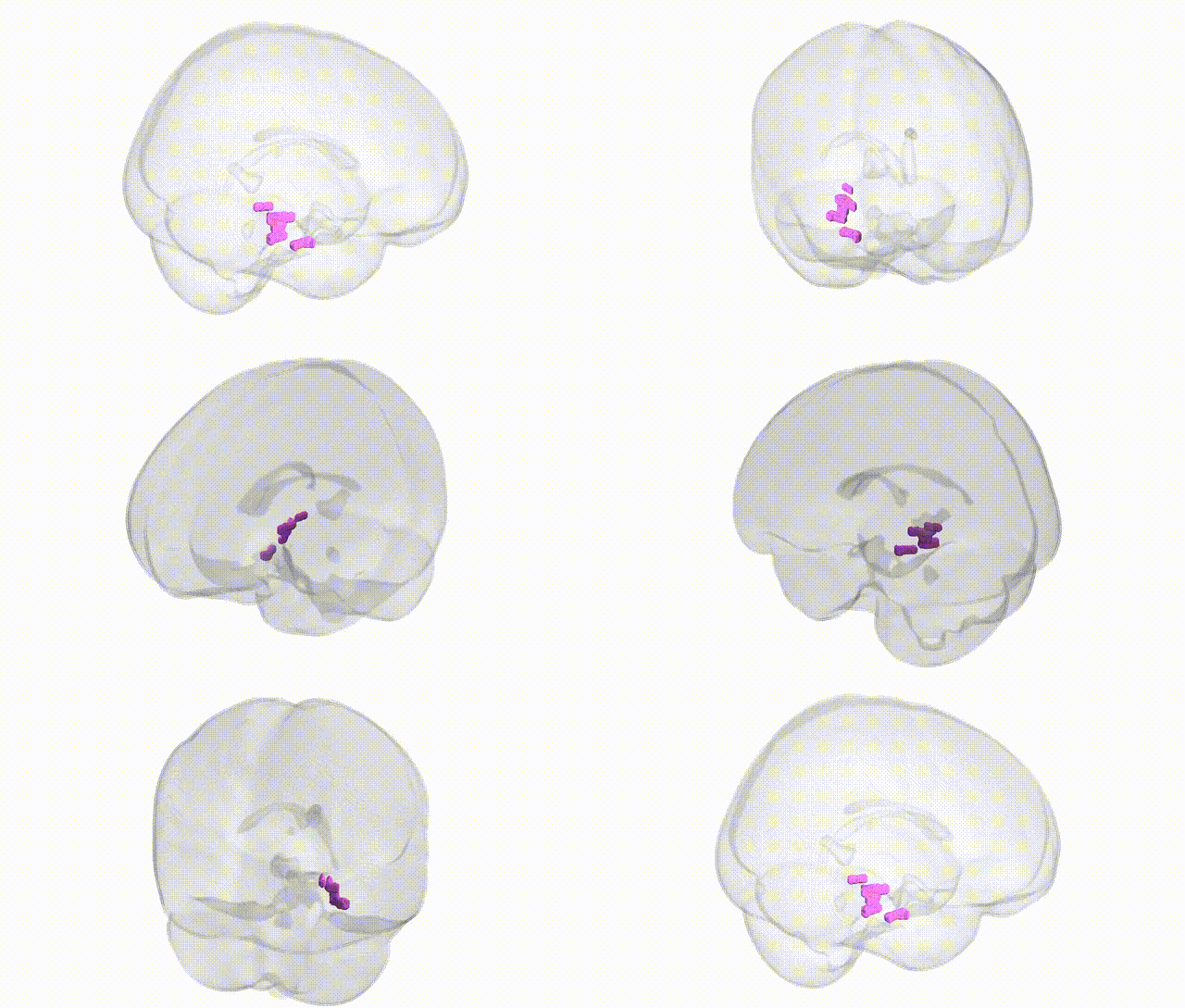
Full Quality Version: Download MP4
Wali Sidiqyar*, Gaurav Rudravaram*, Elyssa M. McMaster, Trent M. Schwartz, Adam M. Saunders, Kurt G. Schilling, Bennett A. Landman "Introducing SPINS: A Shared Public Visualization Library of Neuroanatomical Structures." Medical Imaging with Deep Learning- short paper
This resource is licensed under CC0 1.0 Universal (Public Domain).