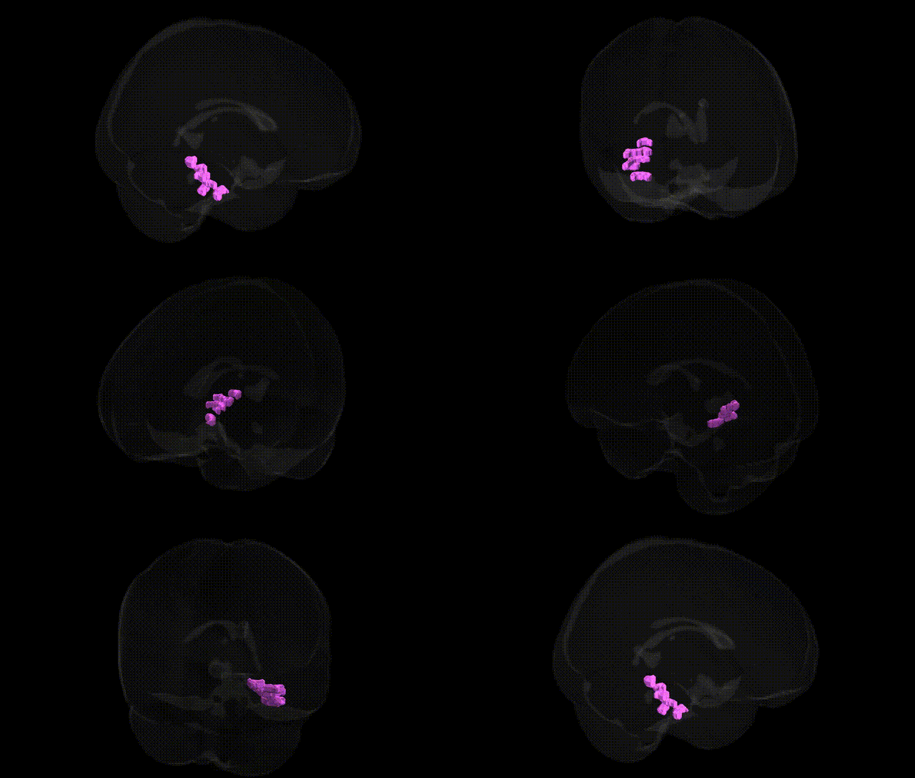
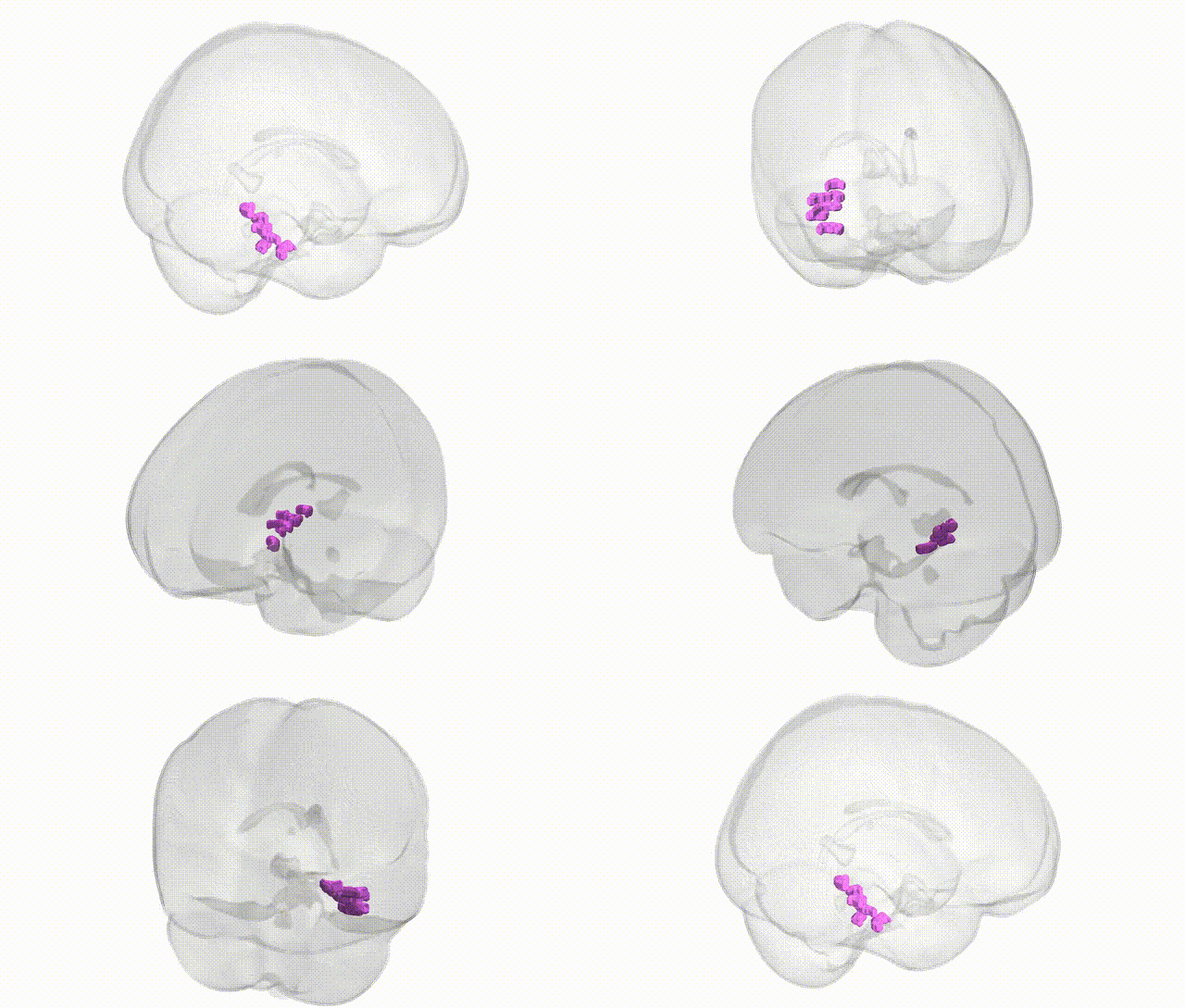
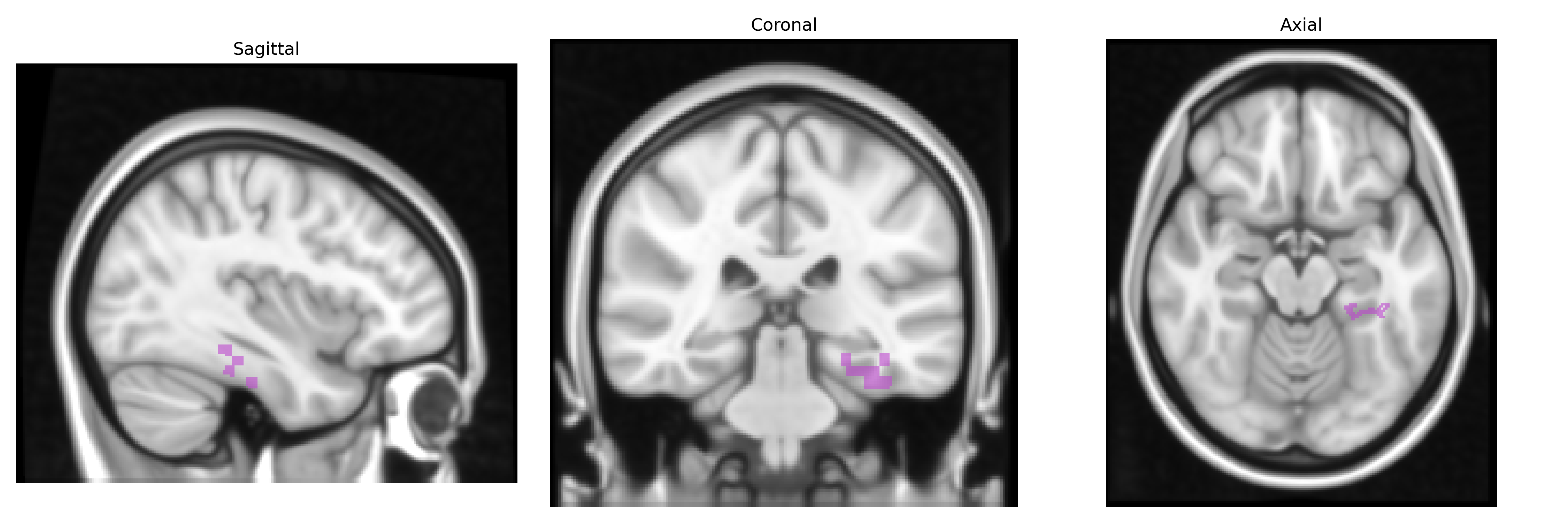
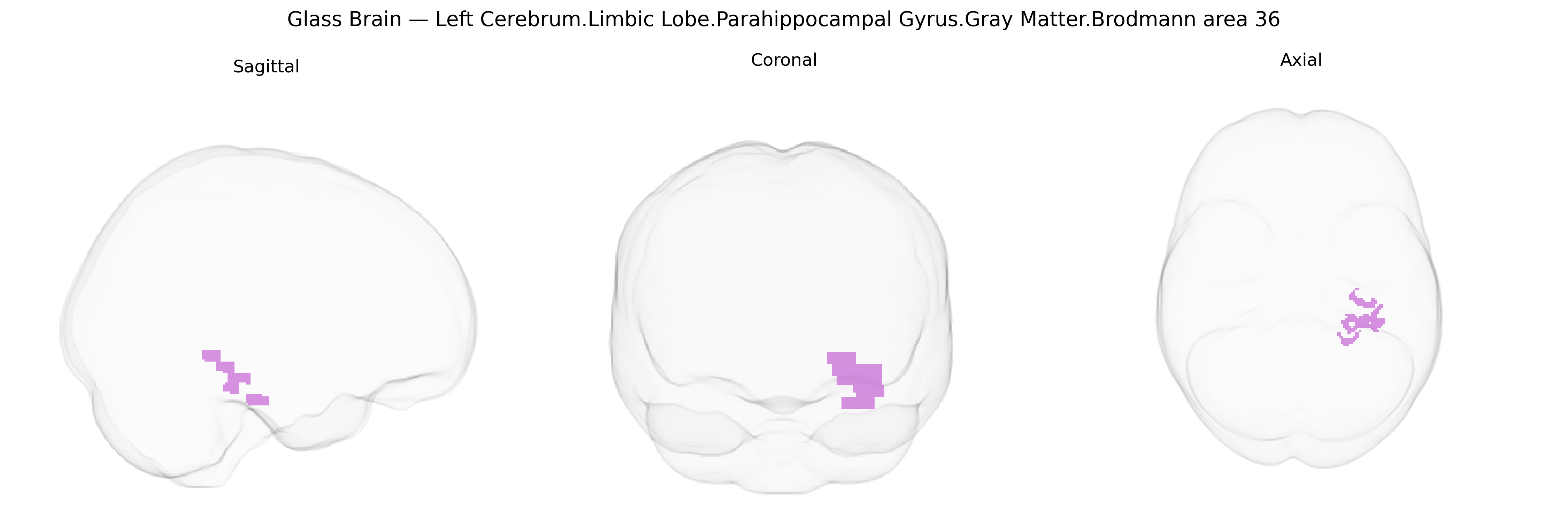
The bilateral Left Cerebrum.Limbic Lobe.Parahippocampal Gyrus.Gray Matter.Brodmann area 36 corresponds to a cytoarchitectonic subdivision of the parahippocampal region involved in higher-order visual association, object and face recognition, and integration of multimodal sensory information with memory processes. Located in the medial temporal lobe, BA36 lies adjacent to the perirhinal cortex and hippocampal formation, forming part of the medial temporal memory system that supports familiarity-based recognition and contributes to semantic memory encoding and retrieval. Neuronal populations in this area receive convergent inputs from ventral visual pathways and project to hippocampal and limbic structures, facilitating the linkage of complex perceptual representations with stored mnemonic traces. There is no direct link for Brodmann area 36; a closely related structure is the Parahippocampal gyrus.
The parahippocampal gyrus (PHG), including Brodmann area 36, shows convergent genetic associations primarily related to memory, emotional processing, and neuropsychiatric and neurodegenerative disorders. Twin and SNP-heritability studies indicate moderate heritability of PHG gray-matter volume and surface measures, with common variants explaining a substantial proportion of inter-individual variance. Large-scale GWAS of brain structure (e.g., ENIGMA and UK Biobank–based studies) have identified multiple loci influencing medial temporal lobe and parahippocampal morphology, including variants in or near genes involved in neurodevelopment, synaptic plasticity, and axon guidance (such as WNT and semaphorin pathway genes), though these often map to broader medial temporal regions rather than BA36 specifically. The PHG has been repeatedly implicated in GWAS and imaging-genetics studies of Alzheimer’s disease, where risk alleles in APOE (especially ε4), CLU, PICALM, BIN1, and other AD loci are associated with atrophy, altered connectivity, or metabolic changes in medial temporal structures encompassing BA36. Schizophrenia and major depression GWAS, combined with imaging endophenotypes, link polygenic risk scores to reduced PHG volume and altered activation, implicating genes in glutamatergic signaling and synaptic organization (including CACNA1C and GRM3 in some studies) in limbic circuitry. In post-traumatic stress disorder and anxiety-related traits, variants in stress-response and neuroplasticity genes such as BDNF (notably Val66Met), FKBP5, and CRHR1 have been associated with structural and functional differences in the parahippocampal/hippocampal complex during fear and context-related memory tasks. Autism spectrum disorder and social-behavior GWAS and rare variant studies suggest that genes involved in synapse formation and chromatin remodeling (e.g., SHANK family genes, NRXN/NRLGN pathways, and some 22q11.2 CNVs) contribute to altered medial temporal and parahippocampal development, although spatial specificity to BA36 is generally inferred from imaging rather than atlas-based localization. Overall, genetic findings point to overlapping sets of neurodevelopmental, synaptic, and neurodegenerative genes influencing structure and function of the parahippocampal gyrus, particularly in relation to context-dependent memory, emotional regulation, and vulnerability to psychiatric and neurodegenerative disorders.
Overview generated by GPT-4o (2026).
Region ID: 129
Hemisphere: bilateral
Atlas: Talairach labels 1mm
Full Quality Version: Download MP4
Full Quality Version: Download MP4
Wali Sidiqyar*, Gaurav Rudravaram*, Elyssa M. McMaster, Trent M. Schwartz, Adam M. Saunders, Kurt G. Schilling, Bennett A. Landman "Introducing SPINS: A Shared Public Visualization Library of Neuroanatomical Structures." Medical Imaging with Deep Learning- short paper
This resource is licensed under CC0 1.0 Universal (Public Domain).