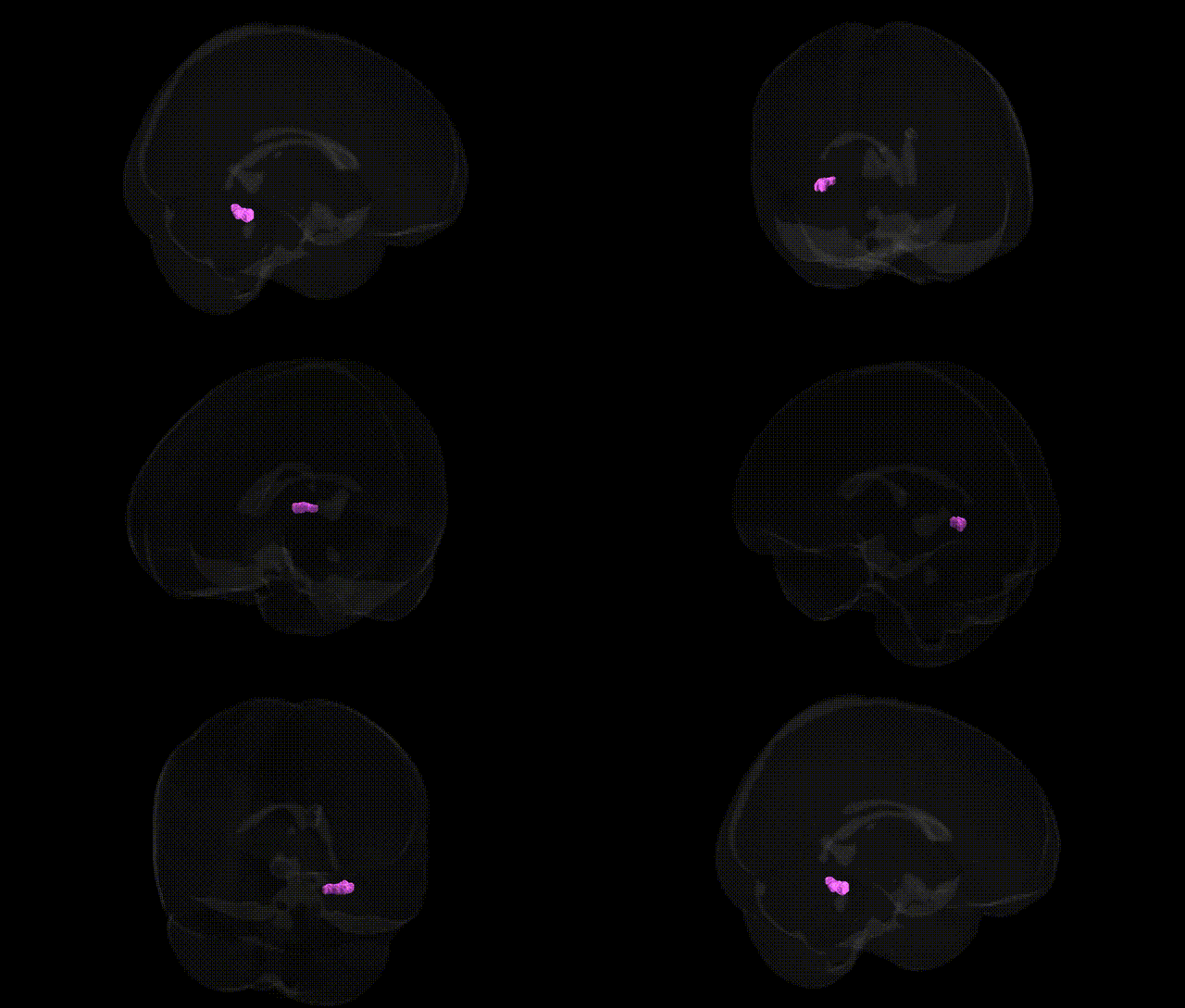
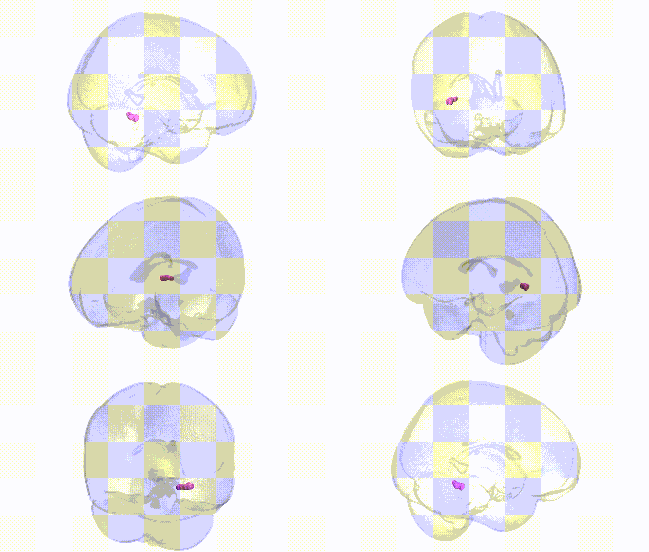
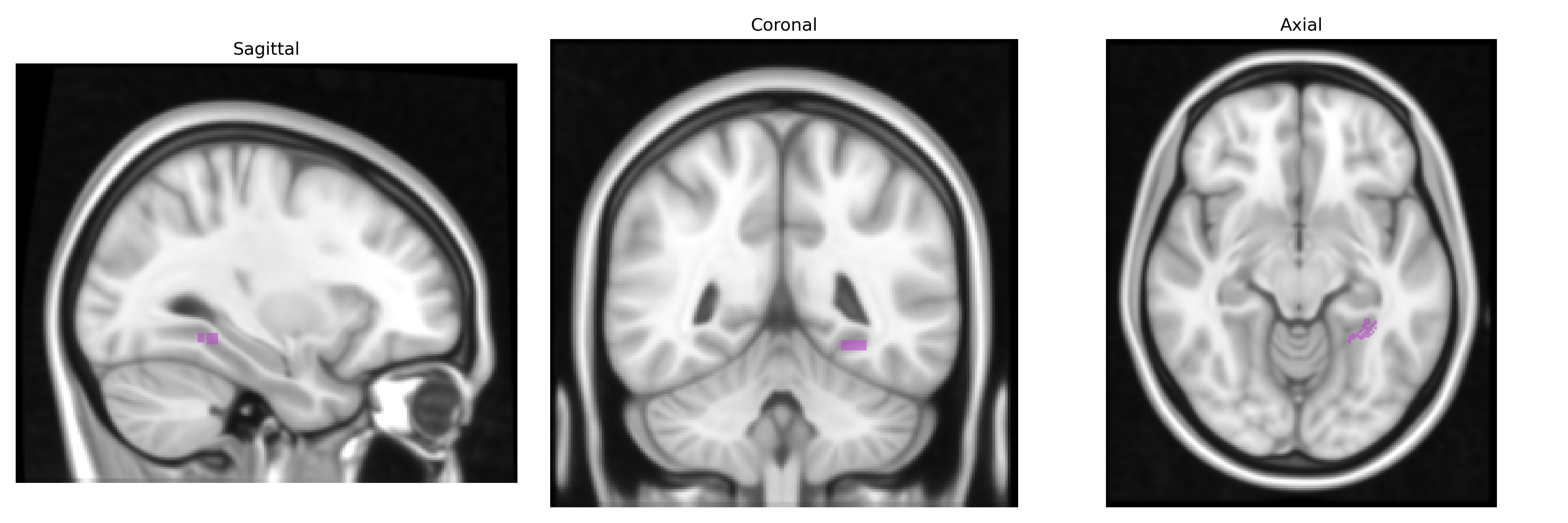
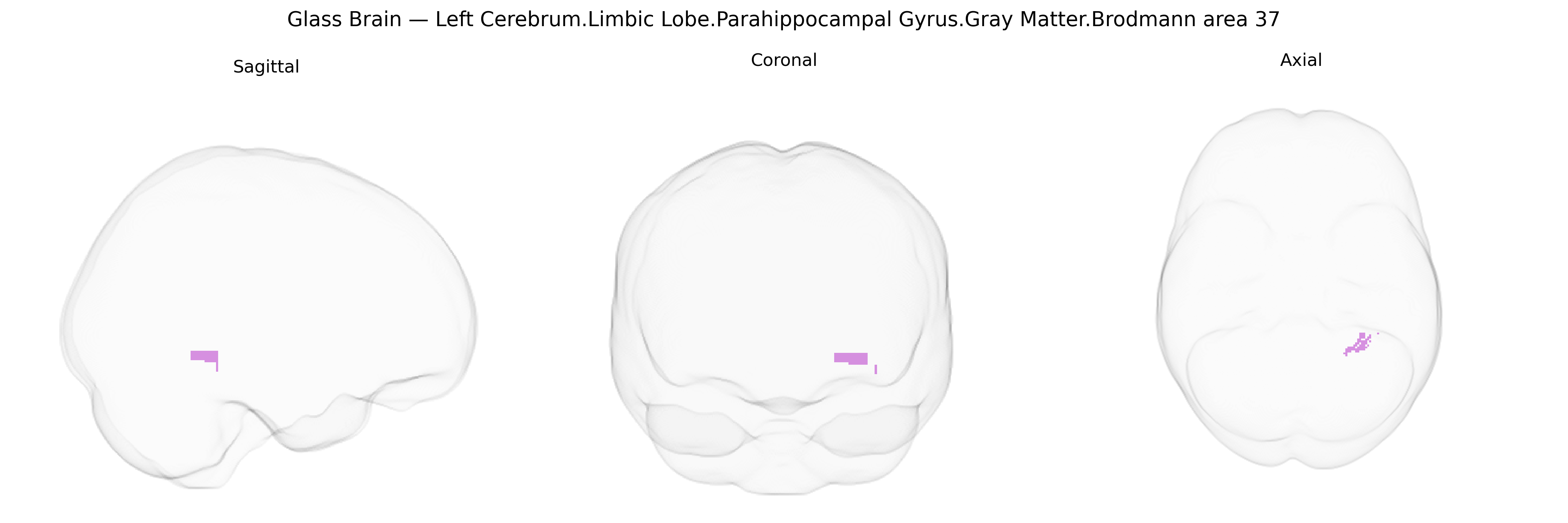
The bilateral Left Cerebrum.Limbic Lobe.Parahippocampal Gyrus.Gray Matter.Brodmann area 37 corresponds primarily to the fusiform and adjacent portions of the parahippocampal region within the inferior temporal and medial temporal cortices, a zone implicated in high-level visual association, semantic processing, and certain aspects of memory. Cytoarchitectonically, Brodmann area 37 is characterized by a well-developed granular layer IV and prominent pyramidal layers III and V, integrating inputs from primary and secondary visual cortices and projecting to temporal, limbic, and frontal association areas. Functionally, this region contributes to the recognition of complex visual stimuli such as faces, objects, and written words, and participates in linking visual patterns with stored semantic and episodic information, thereby supporting perceptual categorization and memory retrieval. Although the Talairach label emphasizes the parahippocampal gyrus and limbic lobe context, BA37 spans a transition between limbic and neocortical territories, positioning it as a critical hub for transforming perceptual input into meaningful, memory-related representations. Brodmann area 37
The parahippocampal gyrus (PHG) in left Brodmann area 37, as defined in the Talairach 1 mm atlas, lies at the interface of medial temporal and ventral occipitotemporal cortices and is repeatedly implicated in genetic studies of memory, emotion, and higher-order visual processing. Twin and SNP-based heritability analyses from large MRI GWAS (e.g., ENIGMA, UK Biobank) show substantial heritability of PHG gray-matter volume and thickness, with common variants in or near genes such as APOE, CLU, PICALM, BIN1, CR1, and SORL1 associating with structural change particularly in Alzheimer’s disease and related dementias, where atrophy of the PHG is an early marker of pathology. GWAS of hippocampal and medial temporal lobe morphology additionally implicate neuronal development and synaptic genes (e.g., BDNF, MAPT region, WNT and TGF-β pathway components), while polygenic risk scores for Alzheimer’s disease, schizophrenia, and major depressive disorder correlate with PHG volume or functional alterations. Structural and functional imaging-genetics work links variation in this region to episodic and contextual memory performance, autobiographical memory, and scene/face processing (often involving fusiform/BA37), with associated loci enriched in genes involved in axon guidance, glutamatergic signaling, and neuroinflammation. Further, GWAS of PTSD, anxiety, and stress-related disorders suggest that risk alleles in HPA-axis and immune-related genes modulate PHG structure and connectivity, consistent with its role in fear and contextual learning, while dysconnectivity and altered activation in left PHG/BA37 have been associated, via genetic imaging studies, with language and reading-related traits and the 22q11.2 and 16p11.2 CNV risk architectures for neurodevelopmental and psychotic disorders.
Overview generated by GPT-4o (2026).
Region ID: 270
Hemisphere: bilateral
Atlas: Talairach labels 1mm
Full Quality Version: Download MP4
Full Quality Version: Download MP4
Wali Sidiqyar*, Gaurav Rudravaram*, Elyssa M. McMaster, Trent M. Schwartz, Adam M. Saunders, Kurt G. Schilling, Bennett A. Landman "Introducing SPINS: A Shared Public Visualization Library of Neuroanatomical Structures." Medical Imaging with Deep Learning- short paper
This resource is licensed under CC0 1.0 Universal (Public Domain).