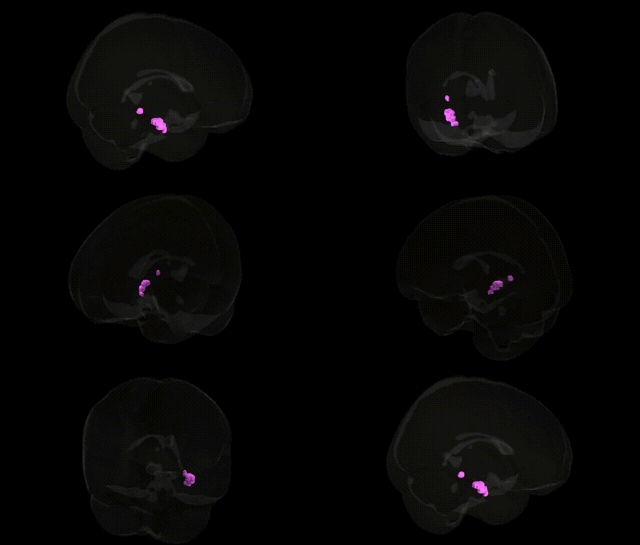
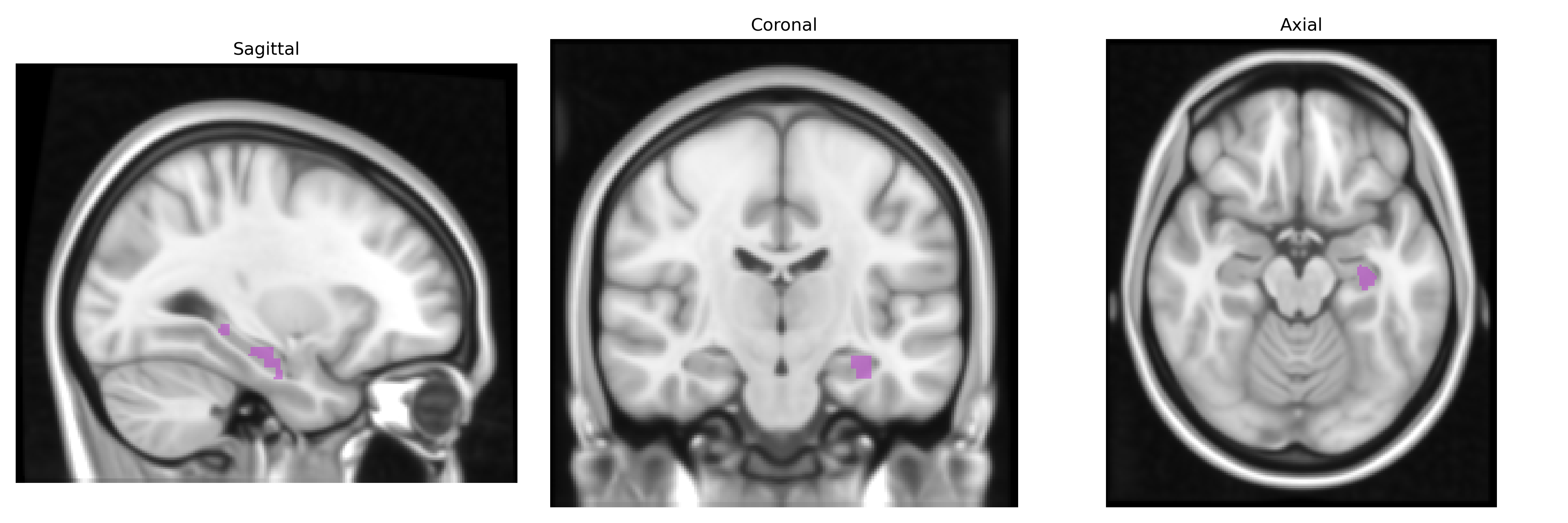
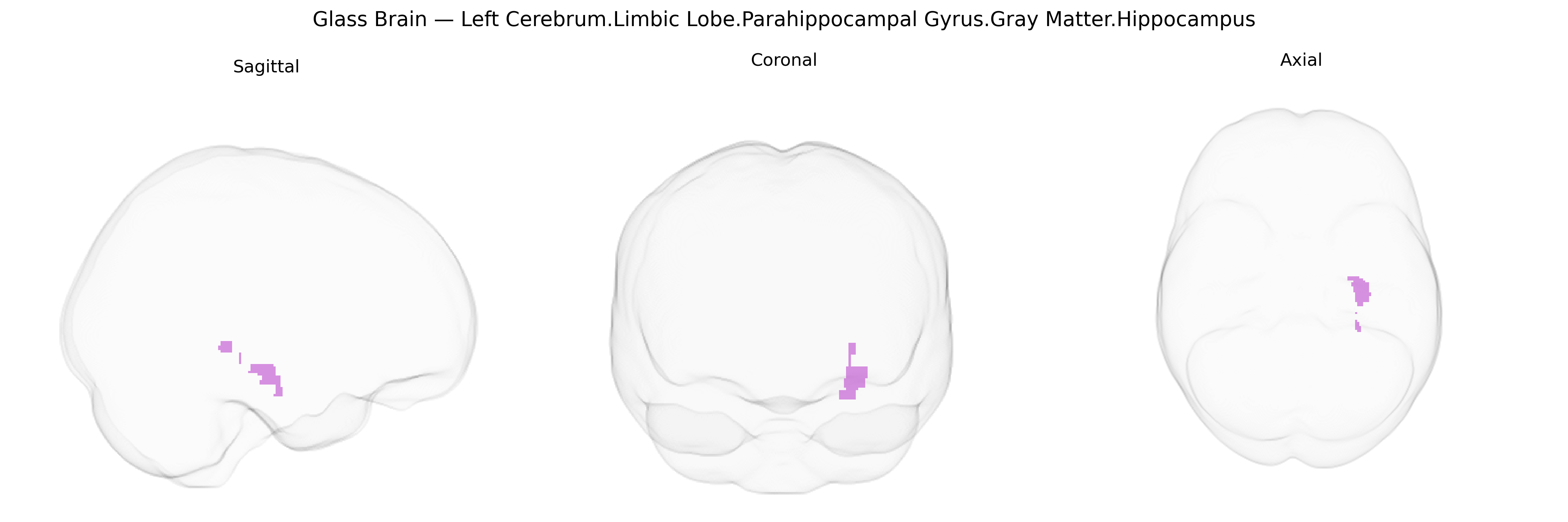
The bilateral Left Cerebrum.Limbic Lobe.Parahippocampal Gyrus.Gray Matter.Hippocampus region corresponds to the hippocampal formation embedded within the medial temporal lobe’s parahippocampal gyrus, forming a core component of the limbic system. Composed primarily of allocortical gray matter (notably the cornu ammonis fields CA1–CA4, dentate gyrus, and subiculum), this structure supports declarative memory encoding, spatial navigation, and contextual association, while also contributing to emotional processing and stress regulation through its extensive connections with the entorhinal cortex, amygdala, thalamus, and prefrontal regions. Its bilateral organization in the left cerebrum is especially important for verbal and contextual memory functions and is highly vulnerable to hypoxic–ischemic injury, temporal lobe epilepsy, and neurodegenerative processes such as those seen in Alzheimer’s disease. Hippocampus
The bilateral parahippocampal gyrus and hippocampus, as defined in Talairach 1 mm atlases, show strong genetic influences and have been central in imaging–genetics and GWAS work on brain structure and neuropsychiatric risk. Twin and SNP-heritability studies (e.g., ENIGMA, UK Biobank) report substantial heritability for hippocampal and parahippocampal gray-matter volume, with common-variant SNP-based heritability often in the 15–35% range, and identify genome-wide significant loci near or within genes such as APOE, SORL1, MAPT, TESC, and others involved in neurodevelopment, synaptic plasticity, and amyloid or tau pathways. Multiple GWAS have linked reduced hippocampal volume—and variation in surrounding parahippocampal cortex—to Alzheimer’s disease and other dementias, with APOE ε4 and numerous late-onset Alzheimer’s risk loci (e.g., CLU, PICALM, BIN1, CR1) associated with atrophy or accelerated volume loss in this region. Polygenic risk scores for Alzheimer’s disease, schizophrenia, major depressive disorder, and bipolar disorder show associations with hippocampal/parahippocampal structural measures, implicating this region in disease vulnerability. Imaging-genetic studies also connect variation in this limbic region to memory performance, general cognitive ability, and stress-related traits, often implicating genes in glutamatergic and GABAergic signaling, neurotrophic pathways (e.g., BDNF), and HPA-axis regulation. Overall, the parahippocampal gyrus and hippocampus emerge as genetically influenced hubs where common and rare variants converge on risk for neurodegenerative and psychiatric disorders, as well as interindividual differences in episodic memory and related cognitive functions.
Overview generated by GPT-4o (2026).
Region ID: 175
Hemisphere: bilateral
Atlas: Talairach labels 1mm
Full Quality Version: Download MP4
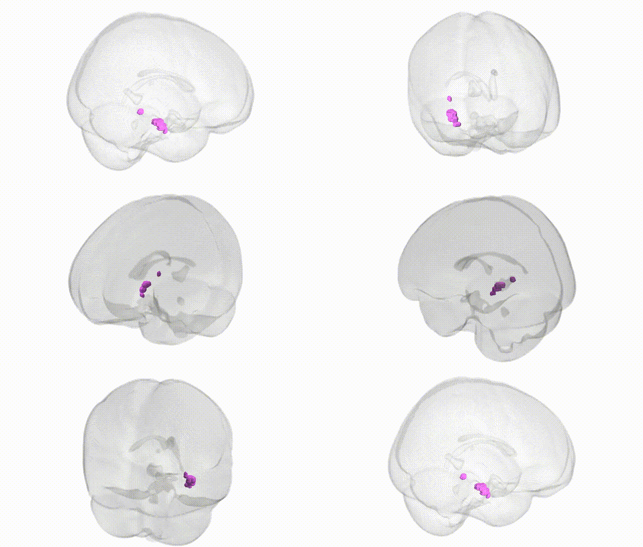
Full Quality Version: Download MP4
Wali Sidiqyar*, Gaurav Rudravaram*, Elyssa M. McMaster, Trent M. Schwartz, Adam M. Saunders, Kurt G. Schilling, Bennett A. Landman "Introducing SPINS: A Shared Public Visualization Library of Neuroanatomical Structures." Medical Imaging with Deep Learning- short paper
This resource is licensed under CC0 1.0 Universal (Public Domain).