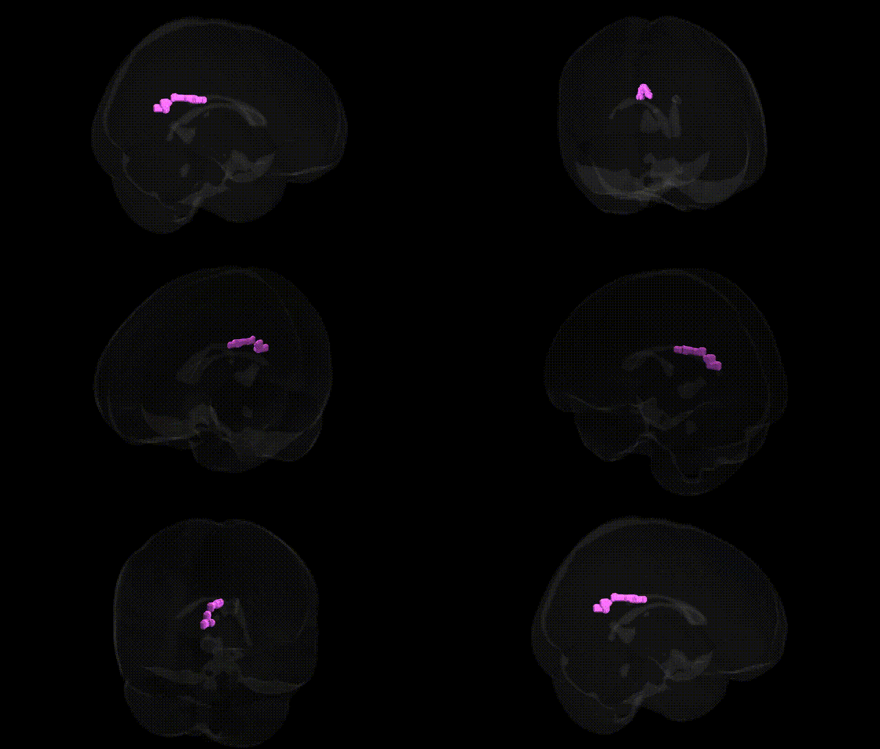
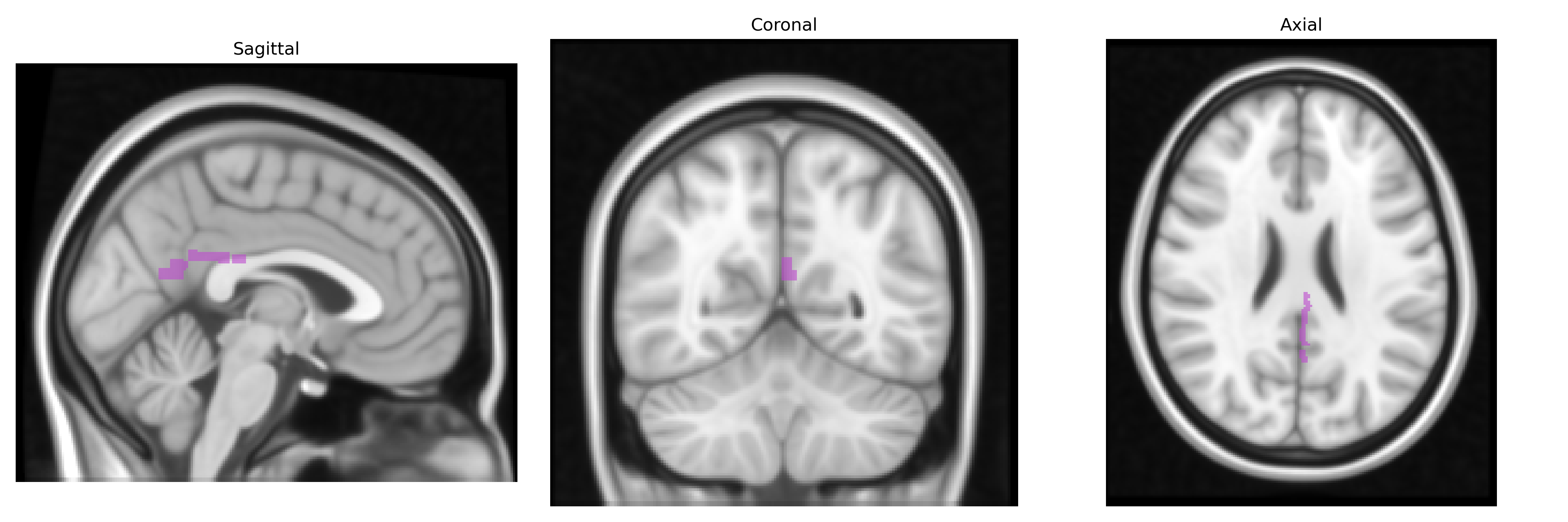
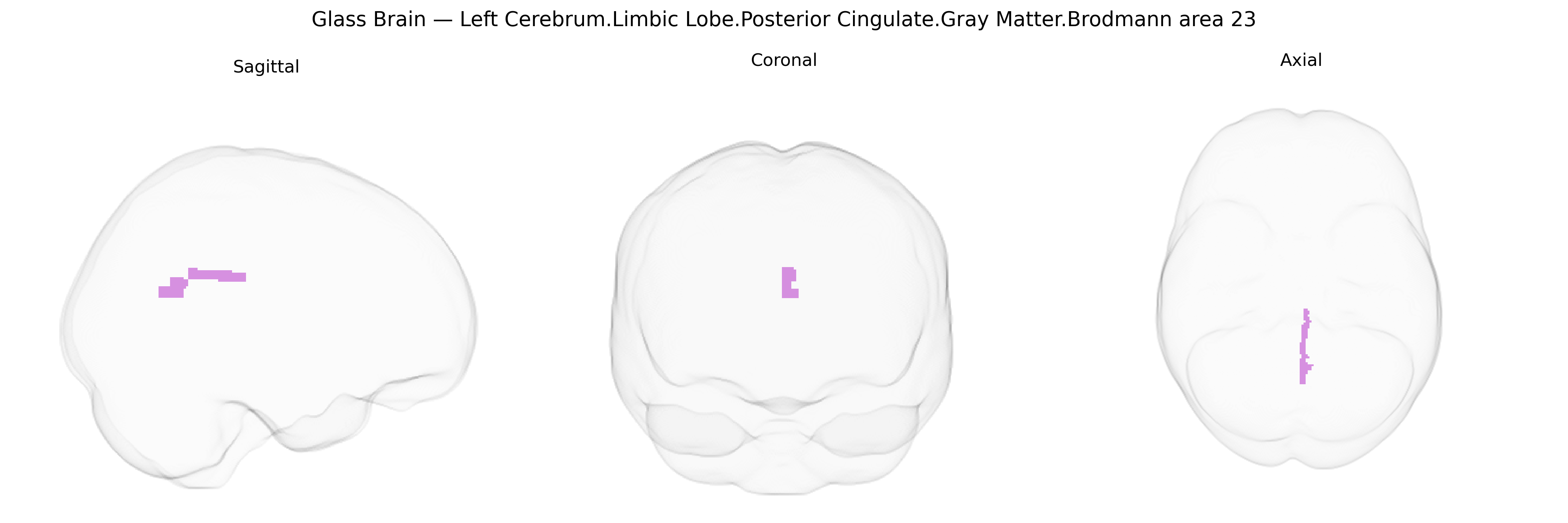
The bilateral Left Cerebrum.Limbic Lobe.Posterior Cingulate.Gray Matter.Brodmann area 23 corresponds to the caudal portion of the posterior cingulate cortex, a limbic-associated cortical field situated on the medial surface of the cerebral hemisphere within the cingulate gyrus. Brodmann area 23 is involved in a range of higher-order integrative functions, including internally directed attention, autobiographical and episodic memory retrieval, visuospatial orientation, and aspects of self-referential and emotional processing, and forms a key node of the default mode network through robust connectivity with the precuneus, hippocampal formation, medial prefrontal cortex, and thalamus. Cytoarchitectonically, it is characterized by a granular isocortical structure distinct from adjacent cingulate areas (e.g., BA24 and BA31), with well-developed layer IV and differentiated pyramidal layers, consistent with its role in multimodal association and limbic-cognitive integration. Brodmann area 23
The bilateral posterior cingulate cortex (PCC; Brodmann area 23 in the limbic lobe) has been repeatedly implicated in imaging–genetics and GWAS studies as a key hub of the default mode network whose structure and function show heritable variation and disease-related genetic associations. Twin and SNP-based heritability analyses indicate moderate to high heritability for PCC gray matter volume, cortical thickness, and functional connectivity, with polygenic influences overlapping those for general cognitive ability, educational attainment, and neuroticism. Large consortia (e.g., ENIGMA, UK Biobank) have identified genome-wide significant associations between common variants in genes involved in synaptic plasticity and neurodevelopment (such as BDNF, APOE, and several glutamatergic and GABAergic pathway genes) and PCC structure or connectivity, including robust effects of the APOE ε4 allele on posterior cingulate atrophy and hypometabolism in Alzheimer’s disease and preclinical stages. PCC alterations linked to genetic risk scores have also been reported in schizophrenia, major depression, and bipolar disorder, where risk alleles and higher polygenic risk scores correlate with reduced PCC volume, disrupted default mode connectivity, or altered task-related deactivation. Additional GWAS of resting-state networks and task fMRI have associated PCC activity patterns with variants influencing immune and microglial pathways, suggesting links between neuroinflammation-related genes and PCC integrity in neurodegenerative and psychiatric conditions.
Overview generated by GPT-4o (2026).
Region ID: 762
Hemisphere: bilateral
Atlas: Talairach labels 1mm
Full Quality Version: Download MP4
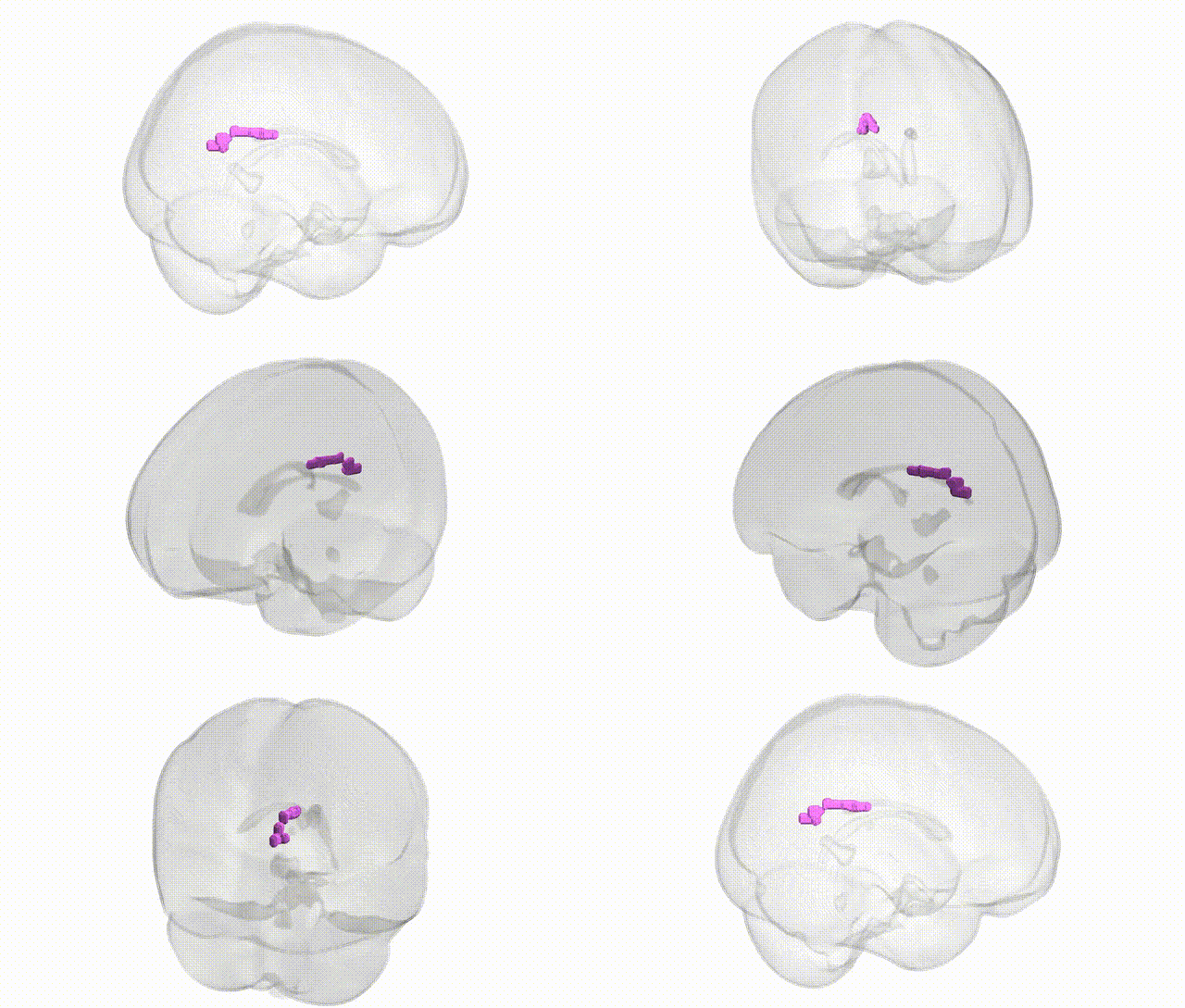
Full Quality Version: Download MP4
Wali Sidiqyar*, Gaurav Rudravaram*, Elyssa M. McMaster, Trent M. Schwartz, Adam M. Saunders, Kurt G. Schilling, Bennett A. Landman "Introducing SPINS: A Shared Public Visualization Library of Neuroanatomical Structures." Medical Imaging with Deep Learning- short paper
This resource is licensed under CC0 1.0 Universal (Public Domain).