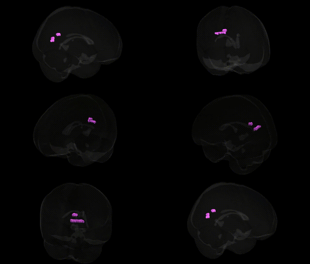
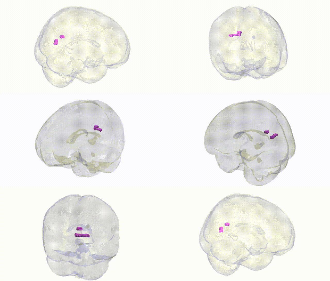
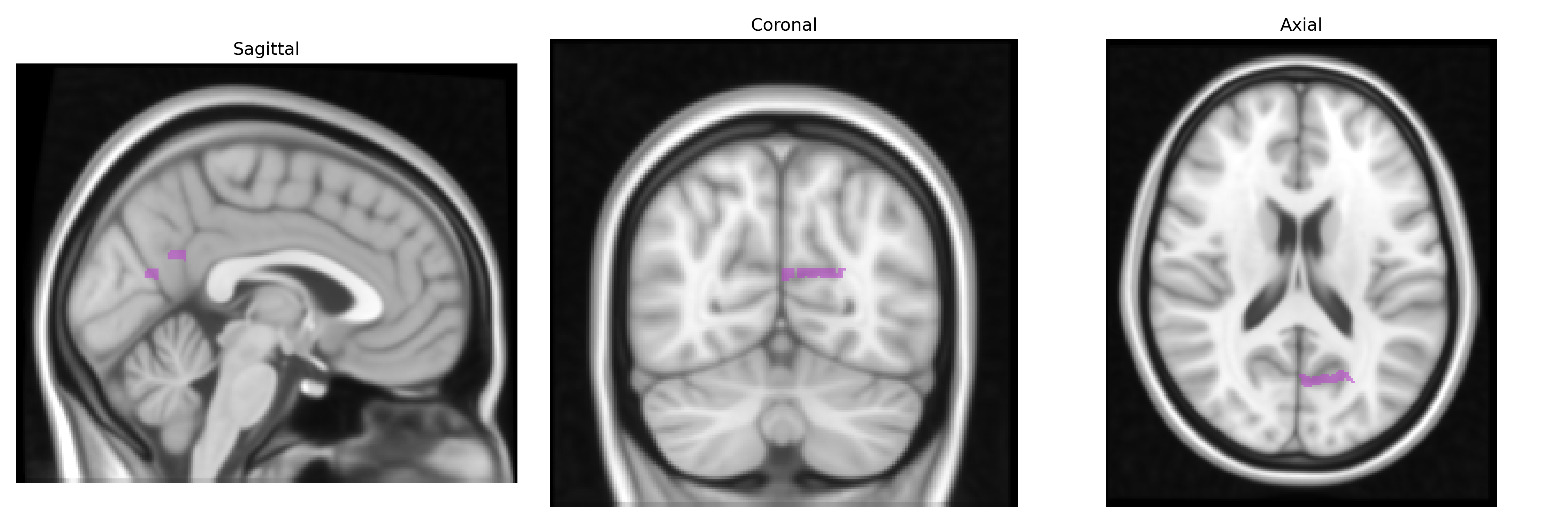
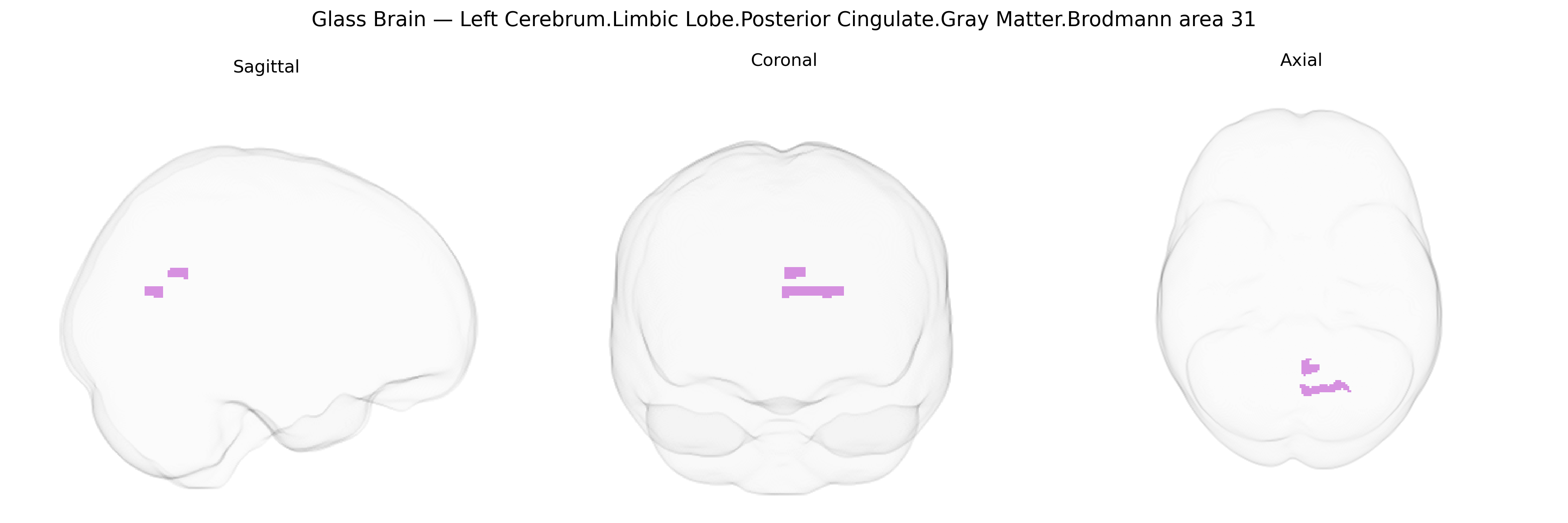
The bilateral Left Cerebrum.Limbic Lobe.Posterior Cingulate.Gray Matter.Brodmann area 31 corresponds to a dorsally located portion of the posterior cingulate cortex, extending along the medial surface of the parietal lobe within the limbic network. Brodmann area 31 is characterized cytoarchitectonically by a granular cortex with well-differentiated layer IV and prominent pyramidal neurons in layers III and V, and functionally participates in episodic memory retrieval, self-referential processing, visuospatial integration, and internal mentation. This region forms a key node of the default mode network, with strong connectivity to the hippocampal formation, precuneus, medial prefrontal cortex, and thalamus, and is implicated in attention to internal states and in neuropathological conditions such as Alzheimer’s disease and other disorders affecting memory and consciousness. Brodmann area 31
Genetic associations involving the bilateral left posterior cingulate cortex (PCC; limbic lobe, Brodmann area 31) identified through GWAS and imaging-genetics studies implicate a range of neuropsychiatric and cognitive phenotypes rather than a single specific locus. Variants in APOE (particularly ε4) and other Alzheimer’s disease risk genes (e.g., CLU, PICALM, BIN1) have been repeatedly associated with altered PCC structure, metabolism, and connectivity, consistent with this region’s early involvement in Alzheimer’s pathology. Polygenic risk for Alzheimer’s disease and for late-life cognitive decline correlates with reduced gray matter volume and disrupted default mode network activity centered on the PCC. Large-scale GWAS of cortical thickness and surface area have identified common variants near genes involved in neurodevelopment, synaptic function, and myelination (e.g., MIR137, HMGA2, WNT and FGF pathway genes) that influence PCC morphology or broader medial parietal–cingulate measures. Psychiatric GWAS and imaging-genetics work link schizophrenia and major depression polygenic risk scores, as well as risk variants in ZNF804A, DISC1, and CACNA1C, to PCC connectivity and default mode network alterations, aligning with its role in self-referential processing and internally directed cognition. Additional associations connect PCC structural and functional variation with genetic liability for traits such as neuroticism, attention-deficit/hyperactivity disorder, and general cognitive ability (g), indicating that common genetic architectures for cognition and affect partly converge on this region.
Overview generated by GPT-4o (2026).
Region ID: 756
Hemisphere: bilateral
Atlas: Talairach labels 1mm
Full Quality Version: Download MP4
Full Quality Version: Download MP4
Wali Sidiqyar*, Gaurav Rudravaram*, Elyssa M. McMaster, Trent M. Schwartz, Adam M. Saunders, Kurt G. Schilling, Bennett A. Landman "Introducing SPINS: A Shared Public Visualization Library of Neuroanatomical Structures." Medical Imaging with Deep Learning- short paper
This resource is licensed under CC0 1.0 Universal (Public Domain).