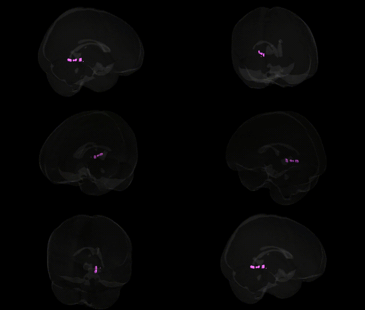
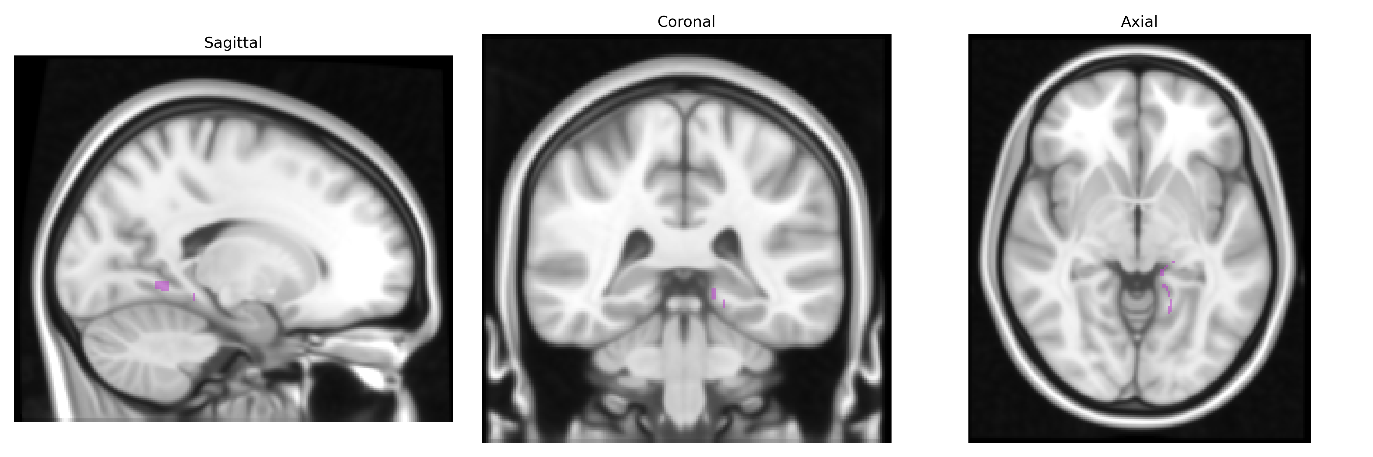
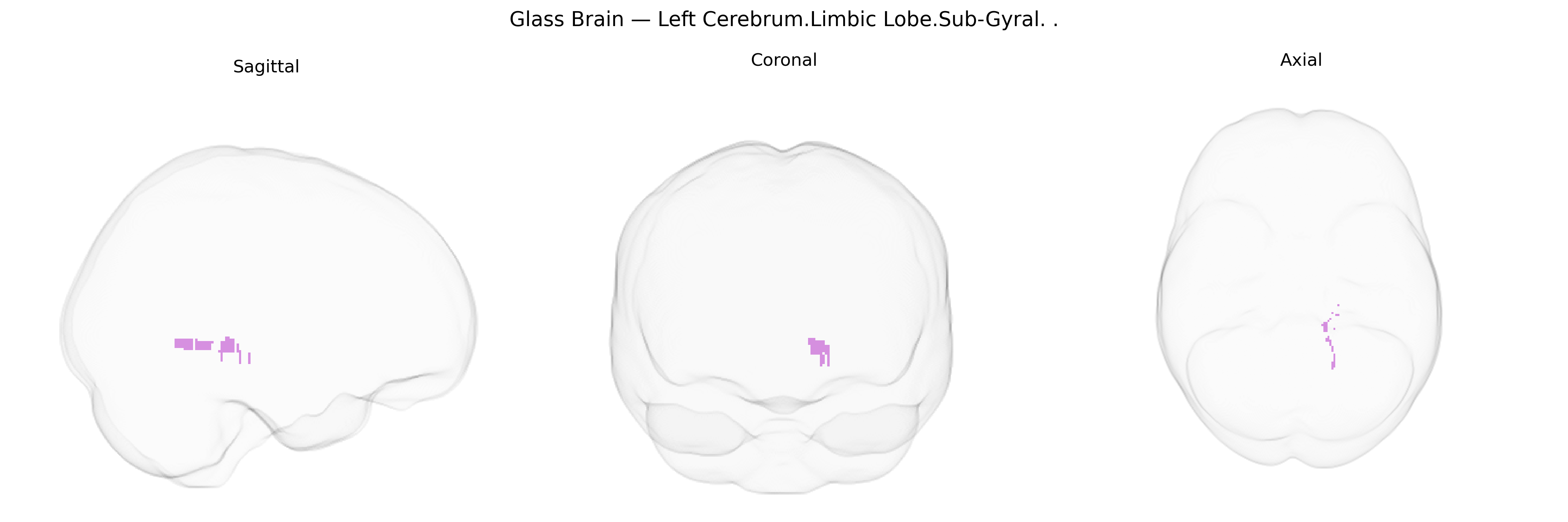
The bilateral Left Cerebrum.Limbic Lobe.Sub-Gyral region in the Talairach 1 mm atlas refers to subcortical white matter located beneath the cortical gyri of the limbic lobe in the left cerebral hemisphere, encompassing fibers that interconnect key limbic structures such as the cingulate gyrus, parahippocampal gyrus, hippocampus, and associated medial temporal and frontal regions. Functionally, this sub-gyral white matter participates in the transmission of information within limbic circuits involved in emotion, memory, affective processing, and autonomic regulation, acting as a conduit between cortical and deeper limbic and paralimbic nuclei. Because “sub-gyral limbic lobe” is an atlas-specific label rather than a single named anatomical structure, there is no direct Wikipedia entry for this exact term; it is typically considered part of the broader Limbic system.
The bilateral left cerebrum limbic sub-gyral region (encompassing deep limbic white and gray matter beneath cingulate and parahippocampal cortices as defined in the Talairach 1 mm atlas) has been implicated in multiple genetics-informed neuroimaging and GWAS studies, largely through broader limbic, cingulate, and medial temporal lobe measures rather than this label alone. Common variants in genes affecting synaptic plasticity, neurodevelopment, and myelination (for example, BDNF, NTRK2, GRIN2B, and neuregulin/ErbB pathway genes) have been associated with limbic volume, cortical thickness, or white-matter integrity in large consortia such as ENIGMA, often overlapping with sub-gyral limbic territories. Polygenic risk scores for major psychiatric disorders—including major depressive disorder, bipolar disorder, schizophrenia, and anxiety—show associations with structural and functional changes in adjacent cingulate and parahippocampal/corticolimbic networks, which extend into sub-gyral limbic areas; analogous associations are seen for neurodegenerative traits such as Alzheimer’s disease risk loci (e.g., APOE ε4 and other GWAS hits) via medial temporal and limbic atrophy patterns. Additional GWAS links involve traits like neuroticism, stress reactivity, and pain sensitivity, where limbic and cingulate sub-gyral regions often emerge in imaging–genetics models connecting specific SNPs or polygenic scores to white-matter microstructure (e.g., fractional anisotropy) and functional connectivity within emotion and memory circuits that traverse this limbic sub-gyral compartment.
Overview generated by GPT-4o (2026).
Region ID: 336
Hemisphere: bilateral
Atlas: Talairach labels 1mm
Full Quality Version: Download MP4
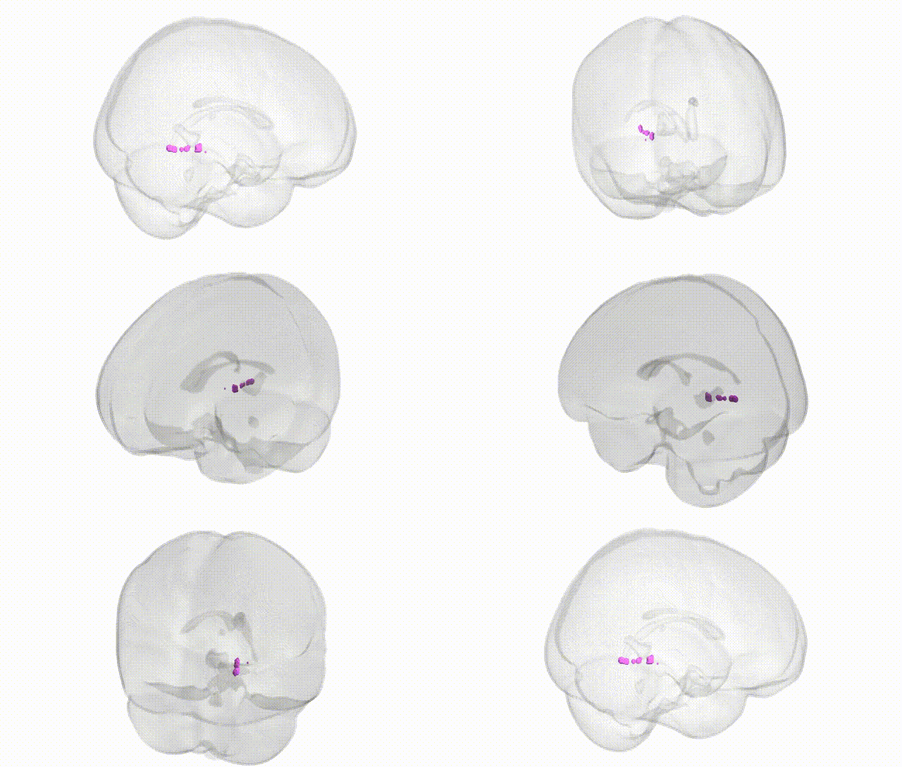
Full Quality Version: Download MP4
Wali Sidiqyar*, Gaurav Rudravaram*, Elyssa M. McMaster, Trent M. Schwartz, Adam M. Saunders, Kurt G. Schilling, Bennett A. Landman "Introducing SPINS: A Shared Public Visualization Library of Neuroanatomical Structures." Medical Imaging with Deep Learning- short paper
This resource is licensed under CC0 1.0 Universal (Public Domain).