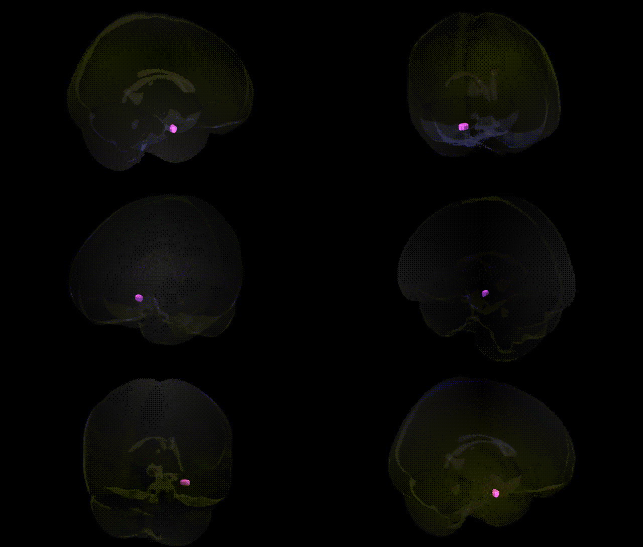
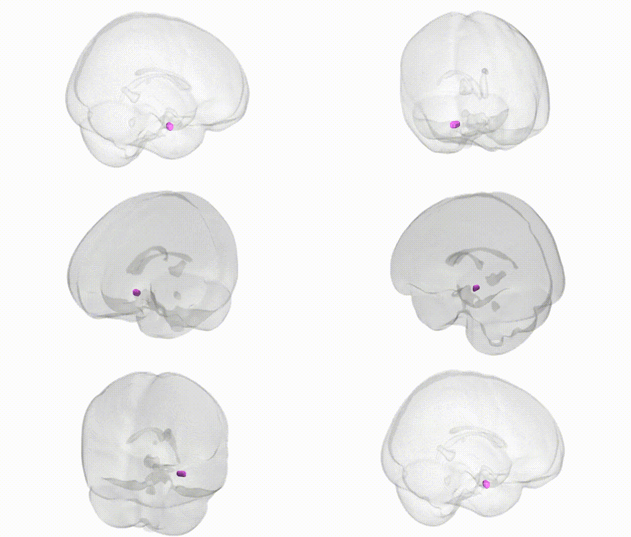
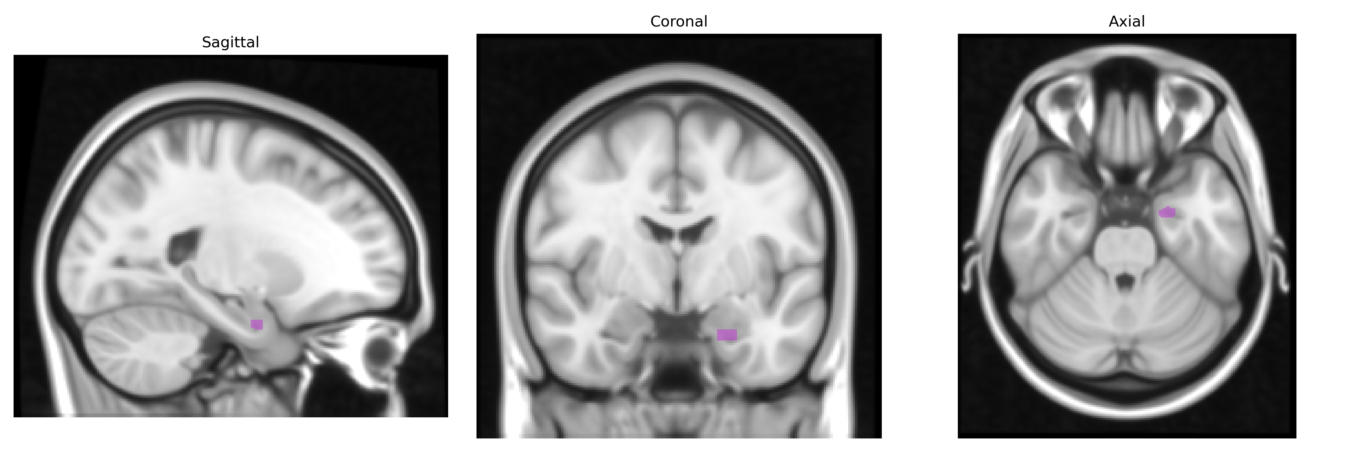
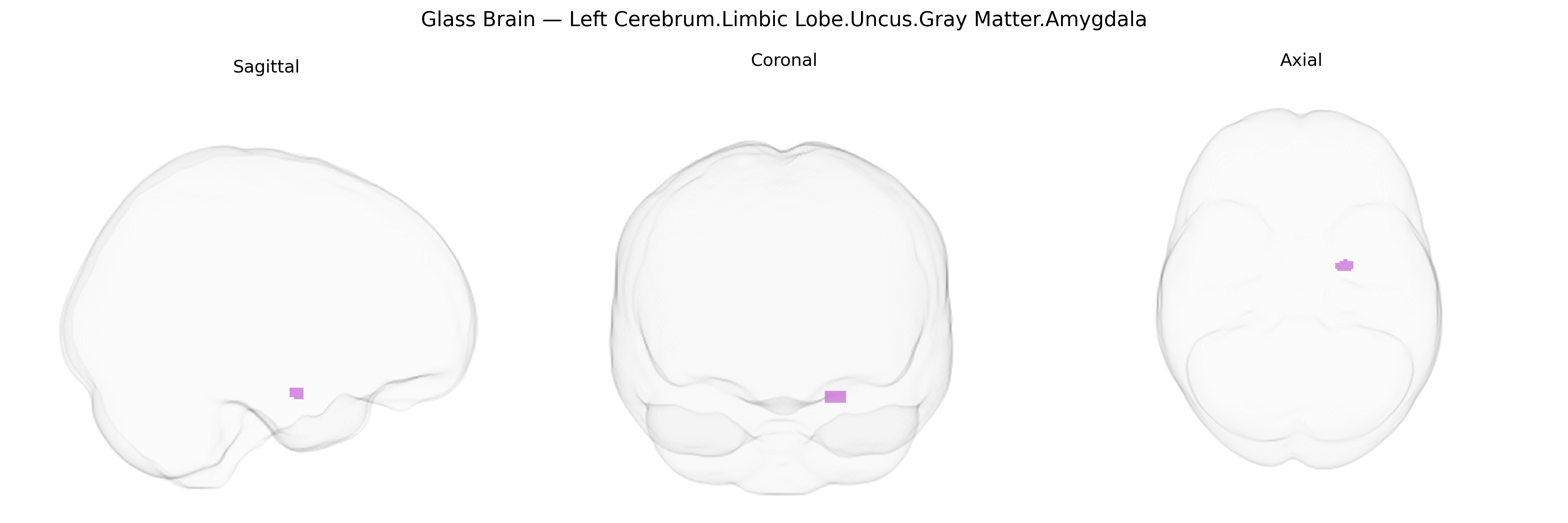
The bilateral Left Cerebrum.Limbic Lobe.Uncus.Gray Matter.Amygdala corresponds to the amygdaloid complex, a collection of nuclei located deep within the medial temporal lobe at the anterior portion of the hippocampal formation, adjacent to the uncus of the parahippocampal gyrus. This predominantly gray matter structure is a key component of the limbic system and is critically involved in processing emotional stimuli, particularly fear and threat detection, as well as in emotional learning, associative memory, and modulation of autonomic and endocrine responses. The amygdala receives multimodal sensory input from cortical and subcortical regions and projects to hypothalamic, brainstem, and prefrontal targets, thereby linking sensory evaluation with visceral, behavioral, and cognitive outputs. Its bilateral organization allows integration of information from both hemispheres, contributing to affective perception, social behavior, and the emotional coloring of memories. Amygdala
The amygdala, located in the uncus of the limbic lobe, shows robust heritability and has been repeatedly implicated in genetic studies of brain structure and neuropsychiatric traits, with GWAS identifying variants influencing its volume and function across multiple loci, including genes involved in neurodevelopment, synaptic signaling, and stress response (e.g., variants near SLC39A8, DRD2, BCL2L1, and other cortical–subcortical patterning genes in large ENIGMA and UK Biobank analyses). Polygenic risk scores for major depressive disorder, schizophrenia, bipolar disorder, autism spectrum disorder, and anxiety-related traits have been associated with altered amygdala volume or reactivity, and specific risk loci for these disorders often converge on pathways affecting glutamatergic and GABAergic transmission, HPA-axis regulation, and neurotrophic signaling, all relevant to amygdala circuitry. GWAS of fear, neuroticism, and stress reactivity indicate shared genetic architecture with amygdala structure and function, while imaging-genetics studies link serotonin transporter (SLC6A4), COMT, and BDNF variants to individual differences in amygdala activation during emotional tasks. Although most large-scale GWAS report amygdala measures at the whole-structure level rather than isolating the uncus, the bilateral limbic amygdala region in the Talairach 1 mm atlas aligns closely with these findings, positioning it as a key node where common genetic variation contributes to risk for mood, anxiety, psychotic, and trauma-related disorders through effects on gray matter morphology and emotional processing networks.
Overview generated by GPT-4o (2026).
Region ID: 181
Hemisphere: bilateral
Atlas: Talairach labels 1mm
Full Quality Version: Download MP4
Full Quality Version: Download MP4
Wali Sidiqyar*, Gaurav Rudravaram*, Elyssa M. McMaster, Trent M. Schwartz, Adam M. Saunders, Kurt G. Schilling, Bennett A. Landman "Introducing SPINS: A Shared Public Visualization Library of Neuroanatomical Structures." Medical Imaging with Deep Learning- short paper
This resource is licensed under CC0 1.0 Universal (Public Domain).