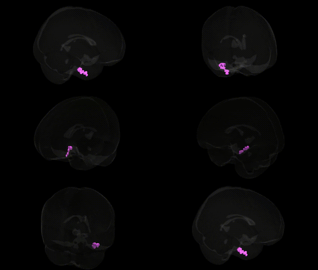
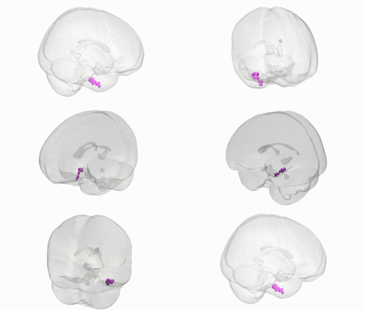
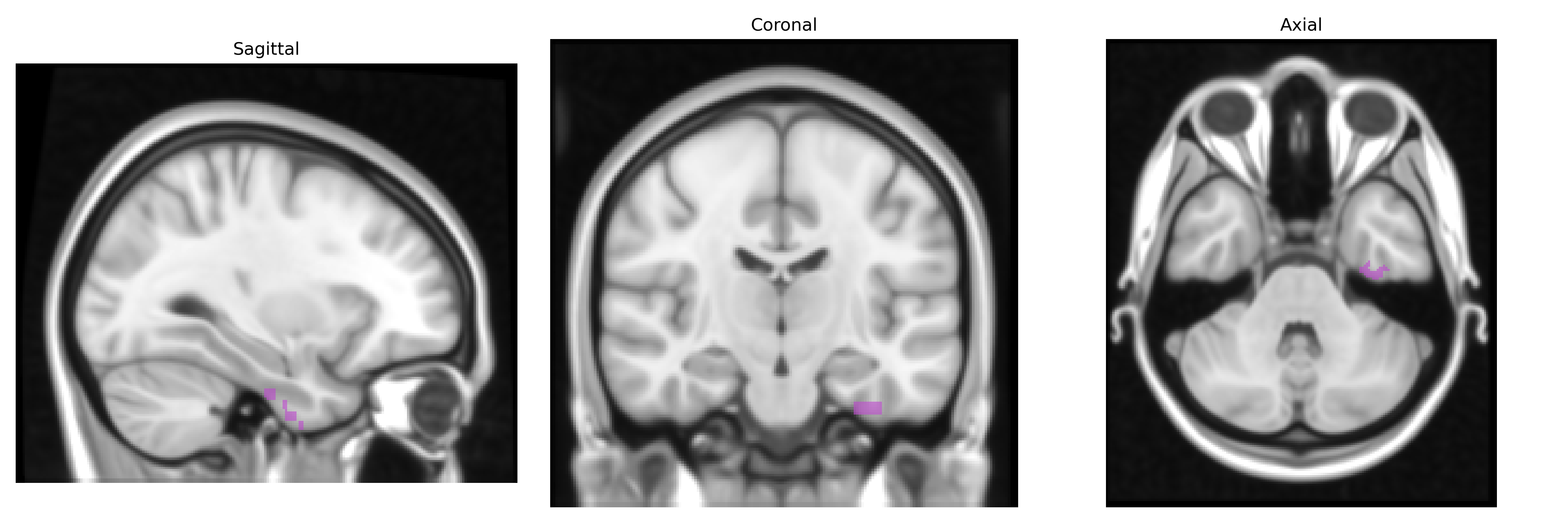
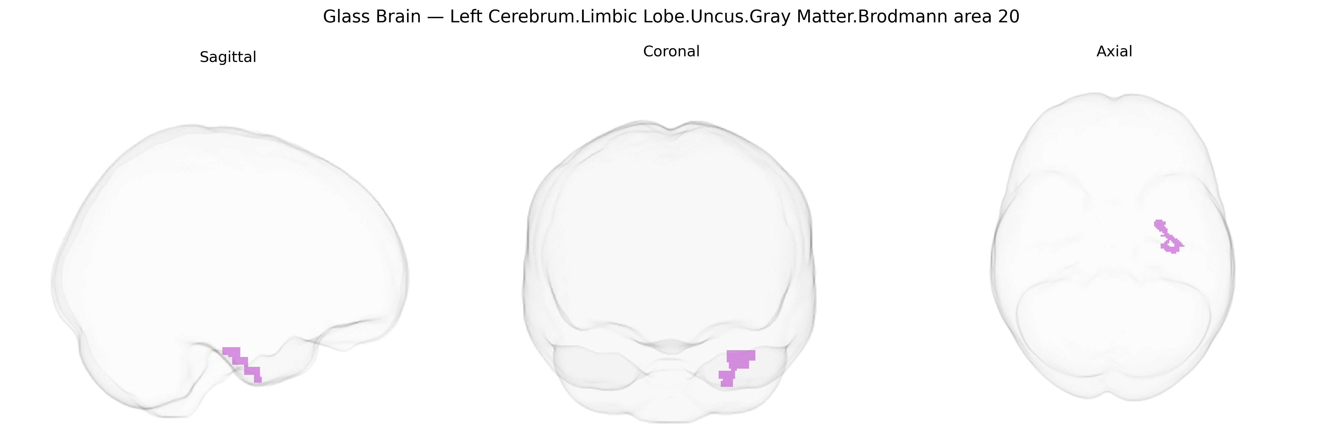
The bilateral Left Cerebrum.Limbic Lobe.Uncus.Gray Matter.Brodmann area 20 refers to gray matter within Brodmann area 20 (BA20) localized in the uncus of the limbic lobe on the left side of the cerebrum, as defined in the Talairach 1 mm atlas. BA20 is part of the inferior temporal cortex and is primarily involved in high‑level visual object recognition, complex feature processing, and contributes to memory and semantic processing through its strong connections with medial temporal lobe structures, including the hippocampal formation and amygdala. In the uncus, this cortex is closely associated with olfactory and emotional processing, and its proximity to hippocampal and parahippocampal regions underlies roles in episodic memory encoding, retrieval, and integration of sensory input with affective and contextual information. There is no direct link for this specific combined region; a related structure is Brodmann area 20.
The bilateral Left Cerebrum Limbic Lobe Uncus gray matter in Brodmann area 20 (anterior medial temporal cortex closely linked with the amygdala and hippocampus) has been implicated in several genetically informed studies, particularly in relation to psychiatric and neurodegenerative disorders and brain-structure GWAS. Variants in genes involved in synaptic plasticity and neurodevelopment (such as BDNF, NTRK2, and RELN) and in glutamatergic and GABAergic signaling have been repeatedly associated with volume, cortical thickness, or functional activity in anterior medial temporal and fusiform/BA20 regions in imaging–genetics work on major depressive disorder, anxiety, schizophrenia, autism spectrum disorder, and obsessive–compulsive symptoms. Large-scale GWAS of subcortical and temporal lobe structures (e.g., ENIGMA and UK Biobank–based studies) have identified common variants near genes such as MSRB3, DPP4, and others influencing temporal and medial temporal gray-matter measures that include or overlap BA20/uncal territory, and these same loci often show pleiotropic associations with Alzheimer’s disease, cognitive performance, and educational attainment. In Alzheimer’s disease and related dementias, risk variants in APOE (particularly ε4), CLU, PICALM, and other AD GWAS genes correlate with accelerated medial/anterior temporal atrophy that extends into uncus/BA20 cortex, while in temporal lobe epilepsy genetic susceptibility loci (including variants affecting excitability and neuroinflammation) have been associated with structural and functional abnormalities in the uncus and adjacent limbic-temporal regions, reflecting the region’s role as a key node in genetically influenced networks for memory, emotion, and seizure propagation.
Overview generated by GPT-4o (2026).
Region ID: 16
Hemisphere: bilateral
Atlas: Talairach labels 1mm
Full Quality Version: Download MP4
Full Quality Version: Download MP4
Wali Sidiqyar*, Gaurav Rudravaram*, Elyssa M. McMaster, Trent M. Schwartz, Adam M. Saunders, Kurt G. Schilling, Bennett A. Landman "Introducing SPINS: A Shared Public Visualization Library of Neuroanatomical Structures." Medical Imaging with Deep Learning- short paper
This resource is licensed under CC0 1.0 Universal (Public Domain).