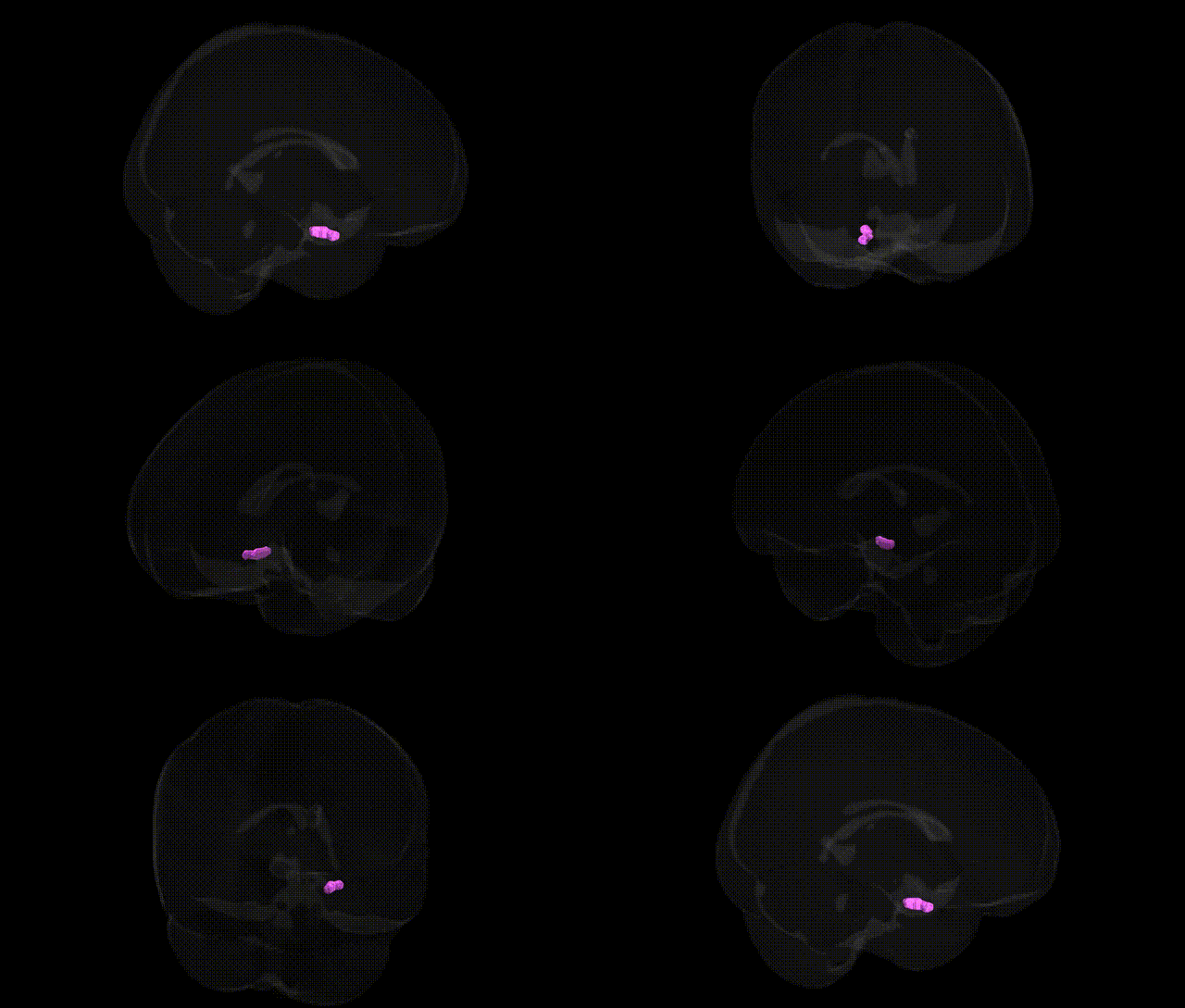
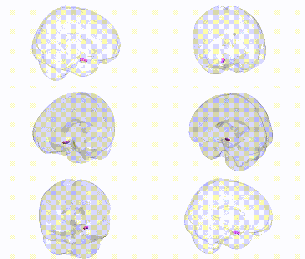
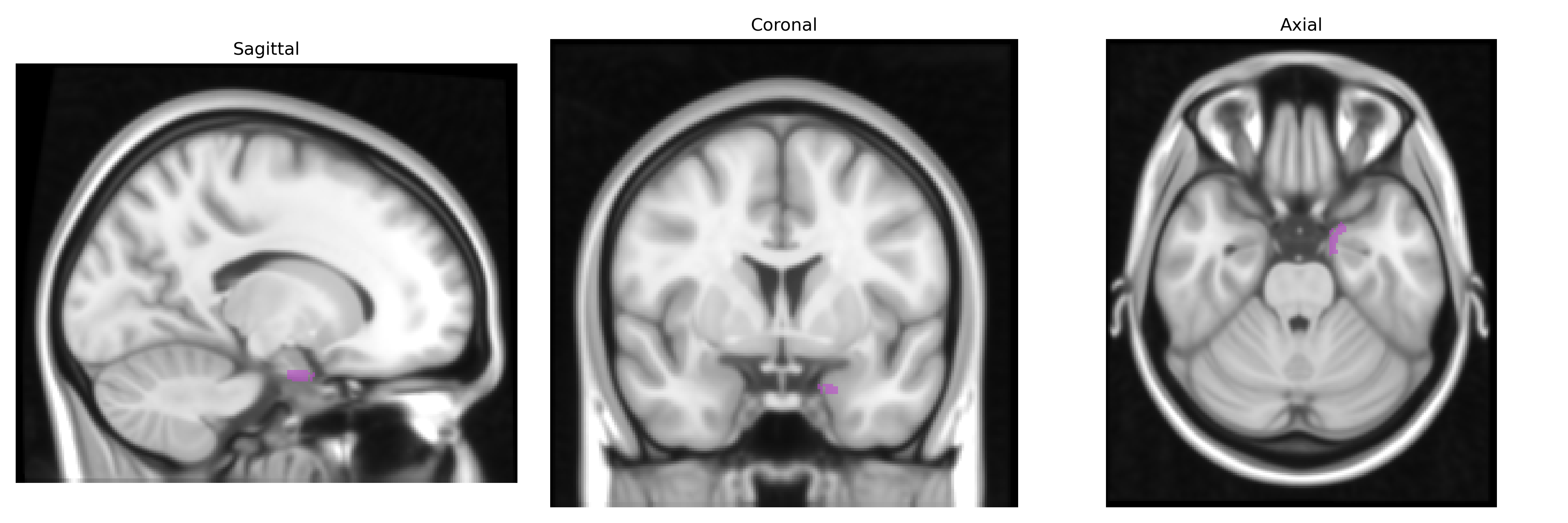
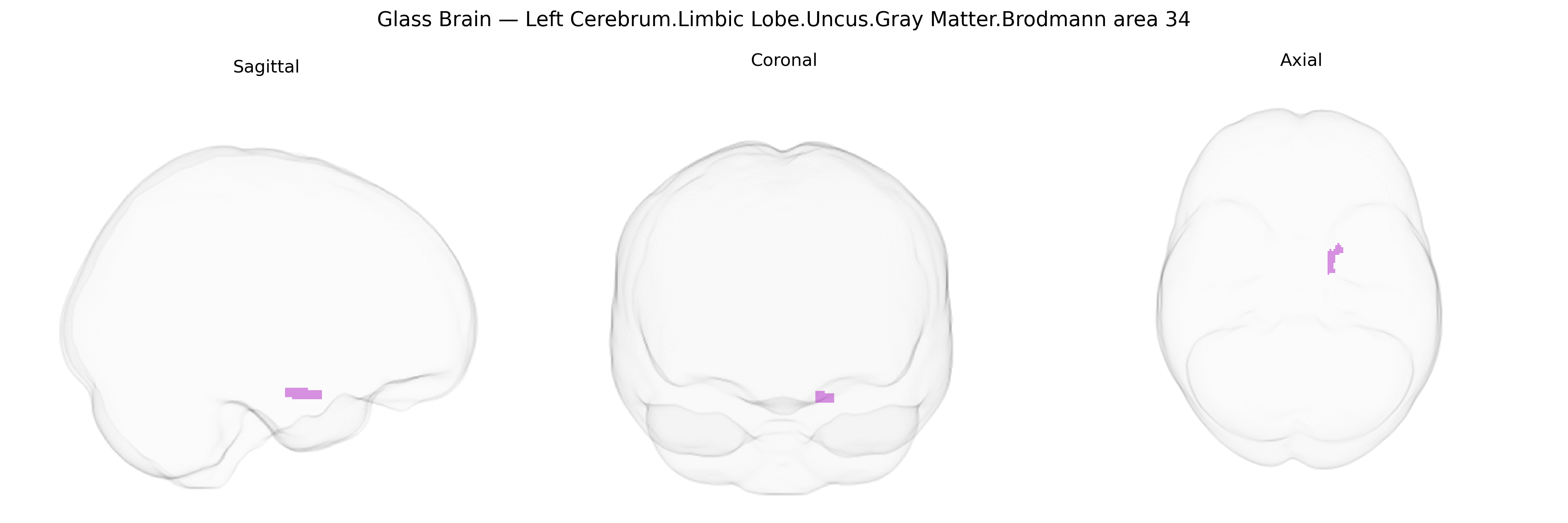
The bilateral Left Cerebrum.Limbic Lobe.Uncus.Gray Matter.Brodmann area 34 corresponds to a portion of the anterior medial temporal lobe situated in the uncus of the parahippocampal gyrus, overlying the amygdaloid complex and closely associated with the hippocampal formation. Brodmann area 34 is typically considered part of the primary olfactory cortex and limbic circuitry, receiving olfactory inputs and contributing to odor perception as well as the integration of olfactory information with emotional and mnemonic processes. Its location in the uncus confers particular clinical significance, as this region is prone to involvement in medial temporal lobe epilepsy and is a key landmark in uncal herniation, where displacement can affect adjacent brainstem structures. There is no direct link for Brodmann area 34 or the uncus alone; a closely related structure with detailed coverage is the Parahippocampal gyrus.
The bilateral Brodmann area 34 (uncus of the limbic lobe) is a medial temporal/entorhinal region heavily implicated in olfactory processing and memory, and genetic associations involving this area largely arise from studies of medial temporal and limbic structures rather than BA34 in isolation. GWAS of hippocampal and entorhinal cortical volume and thickness have identified common variants in genes such as KIBRA (WWC1), APOE, TOMM40, CLU, and CR1 that influence medial temporal morphology and Alzheimer’s disease risk, with neuroimaging genetics studies frequently showing volumetric and functional alterations extending into adjacent uncus/BA34 territory. APOE ε4 status has been repeatedly associated with reduced medial temporal gray matter, altered activation, and accelerated atrophy in prodromal Alzheimer’s disease, while other GWAS of limbic and temporal lobe measures have implicated loci involved in synaptic function and neurodevelopment, including genes like BDNF, GRIN2B, and genes within the 22q11.2 region, which also carry susceptibility to schizophrenia and related cognitive and structural changes in medial temporal cortices. Variants in serotonin (e.g., HTR1A, HTR2A) and glutamate-related genes have been linked to anxiety, depression, and post-traumatic stress disorder with convergent imaging evidence of abnormal activation and connectivity in uncus/BA34 and adjacent limbic regions. Olfactory deficits, an early feature in neurodegenerative and psychiatric disorders that involve BA34, have genetic contributions via loci affecting olfactory receptor gene clusters and neurodegeneration risk genes (e.g., SNCA, GBA) in Parkinson’s disease, with imaging studies showing engagement of uncus and entorhinal cortex. Overall, genetic findings for BA34 primarily reflect broader limbic and medial temporal system associations with Alzheimer’s disease, schizophrenia, mood and anxiety disorders, and neurodegenerative traits, rather than region-specific GWAS focused solely on the uncus.
Overview generated by GPT-4o (2026).
Region ID: 179
Hemisphere: bilateral
Atlas: Talairach labels 1mm
Full Quality Version: Download MP4
Full Quality Version: Download MP4
Wali Sidiqyar*, Gaurav Rudravaram*, Elyssa M. McMaster, Trent M. Schwartz, Adam M. Saunders, Kurt G. Schilling, Bennett A. Landman "Introducing SPINS: A Shared Public Visualization Library of Neuroanatomical Structures." Medical Imaging with Deep Learning- short paper
This resource is licensed under CC0 1.0 Universal (Public Domain).