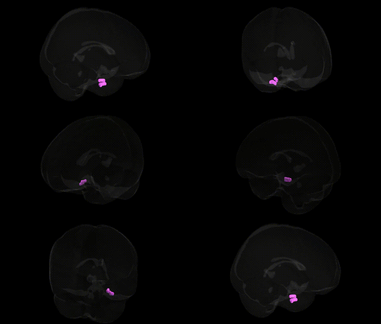
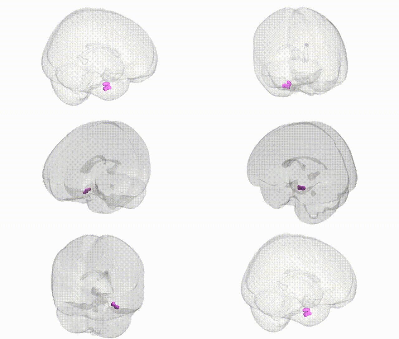
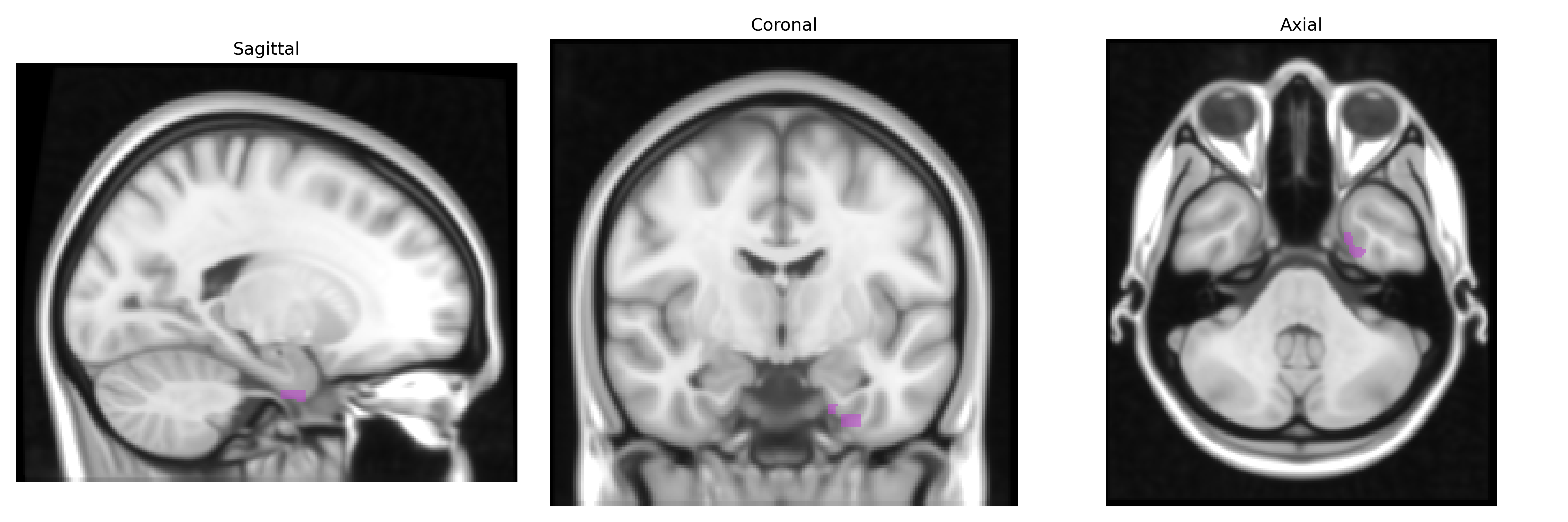
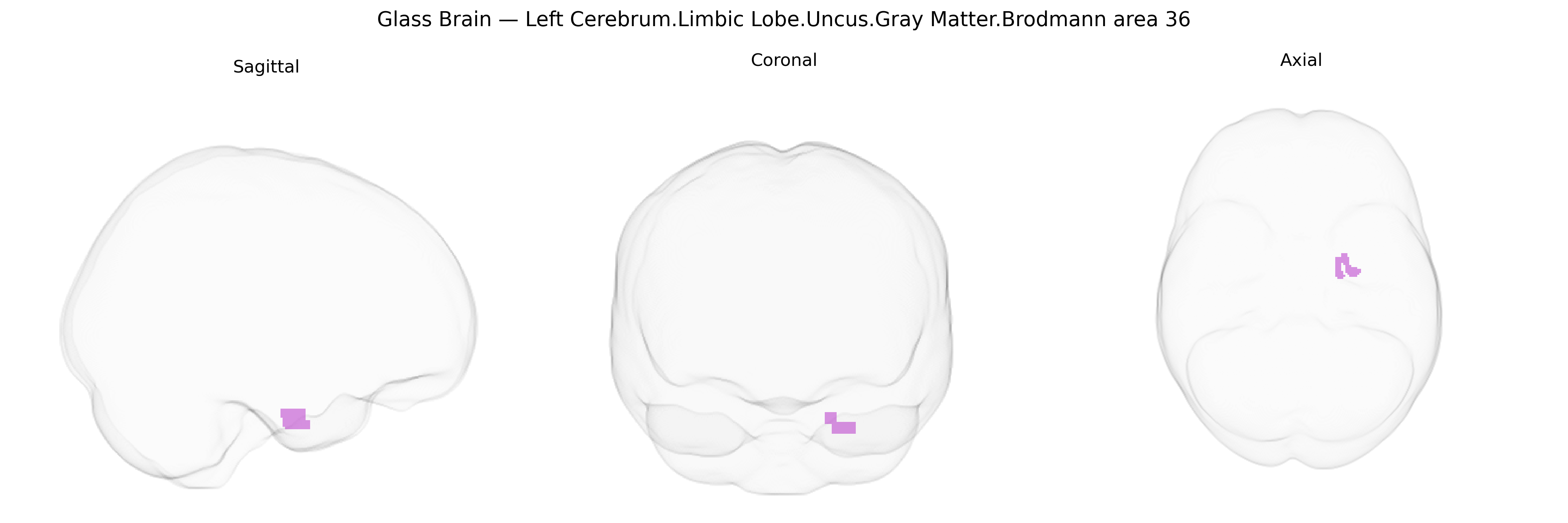
Bilateral Left Cerebrum.Limbic Lobe.Uncus.Gray Matter.Brodmann area 36 corresponds to cortical tissue within the parahippocampal portion of the limbic lobe, specifically involving the perirhinal cortex mapped as Brodmann area 36 in the medial temporal lobe, adjacent to the uncus. This region is implicated in higher-order visual association and memory processes, including object recognition, familiarity judgments, and integration of multimodal sensory information with mnemonic representations. Functionally, it participates in the medial temporal lobe memory system, interacting with the hippocampus and amygdala to support declarative memory, emotional modulation of memory, and aspects of semantic processing. Its cytoarchitecture is characterized by a layered neocortical structure transitioning toward allocortical regions of the parahippocampal gyrus. There is no direct Wikipedia article for “Brodmann area 36”; a closely related structure is the Parahippocampal gyrus.
The left Brodmann area 36 (BA36) in the uncus/limbic lobe, encompassing anterior parahippocampal and perirhinal cortex, has been implicated in several genetic and GWAS-based associations, primarily via imaging-genetics and psychiatric/neurologic trait studies. Structural and functional variation in this region has been linked to common risk variants for Alzheimer’s disease (notably APOE ε4, CLU, BIN1, and SORL1), which show associations with parahippocampal and perirhinal atrophy and altered activation during memory tasks, as well as with polygenic risk scores for Alzheimer’s and related dementias. Large neuroimaging GWAS consortia (e.g., ENIGMA, UK Biobank–based studies) have reported significant SNP heritability and loci affecting cortical thickness or surface area in medial temporal regions that include BA36, with variants in genes related to neurodevelopment (e.g., WNT pathway genes), synaptic function, and axonal guidance, though often at the level of broader parahippocampal or temporal lobe ROIs rather than BA36 alone. Psychiatric GWAS and imaging-genetics work have linked this region’s gray matter volume and connectivity with polygenic risk for schizophrenia, major depression, and autism spectrum disorder, reflecting its role in memory, contextual processing, and emotional evaluation. Additional associations include genetic influences on recognition memory performance and face/object memory tasks that critically depend on perirhinal cortex, as well as variants tied to temporal lobe epilepsy and medial temporal sclerosis, where BA36/uncal regions often show genetically influenced susceptibility to sclerosis and volume loss.
Overview generated by GPT-4o (2026).
Region ID: 75
Hemisphere: bilateral
Atlas: Talairach labels 1mm
Full Quality Version: Download MP4
Full Quality Version: Download MP4
Wali Sidiqyar*, Gaurav Rudravaram*, Elyssa M. McMaster, Trent M. Schwartz, Adam M. Saunders, Kurt G. Schilling, Bennett A. Landman "Introducing SPINS: A Shared Public Visualization Library of Neuroanatomical Structures." Medical Imaging with Deep Learning- short paper
This resource is licensed under CC0 1.0 Universal (Public Domain).