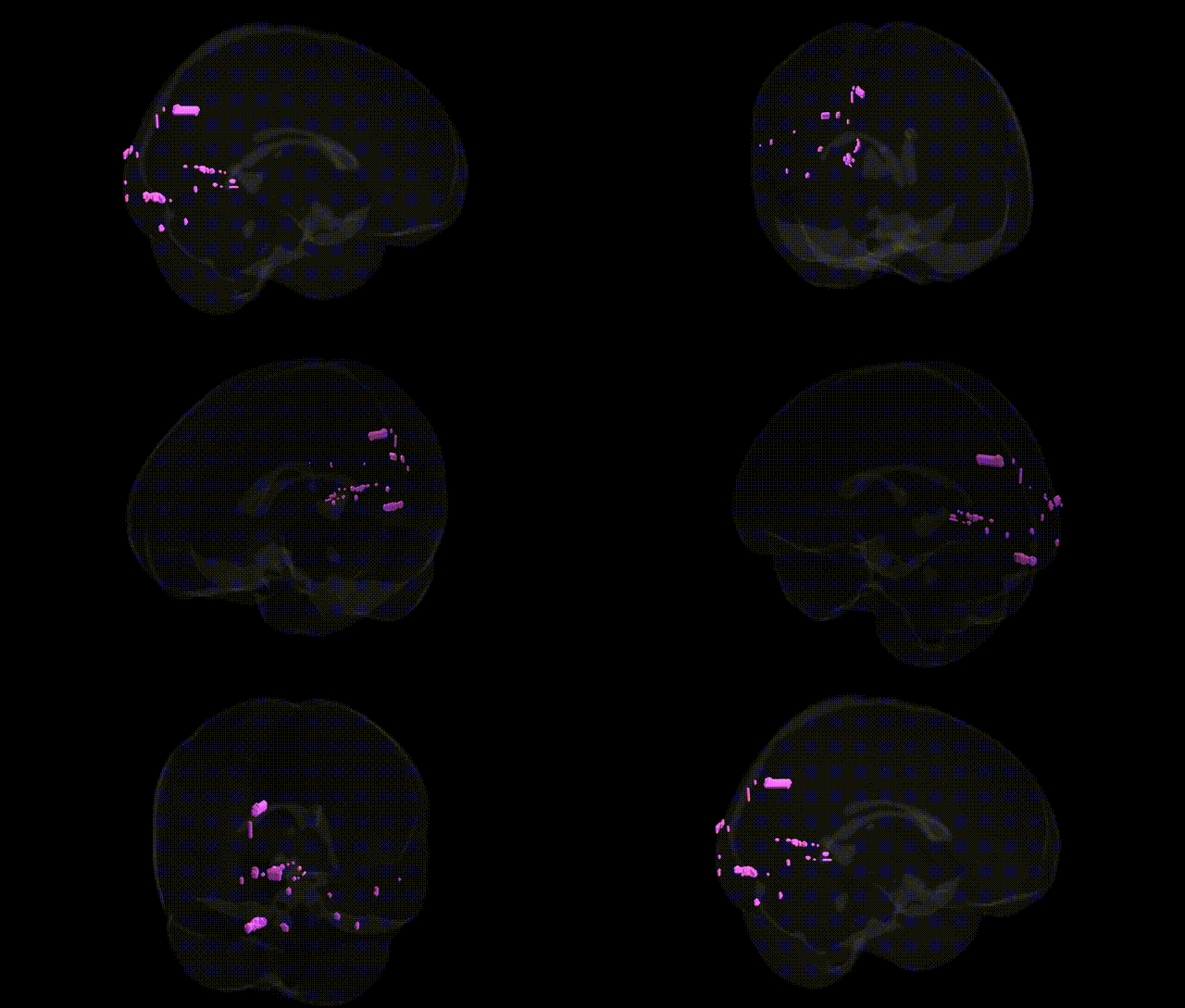
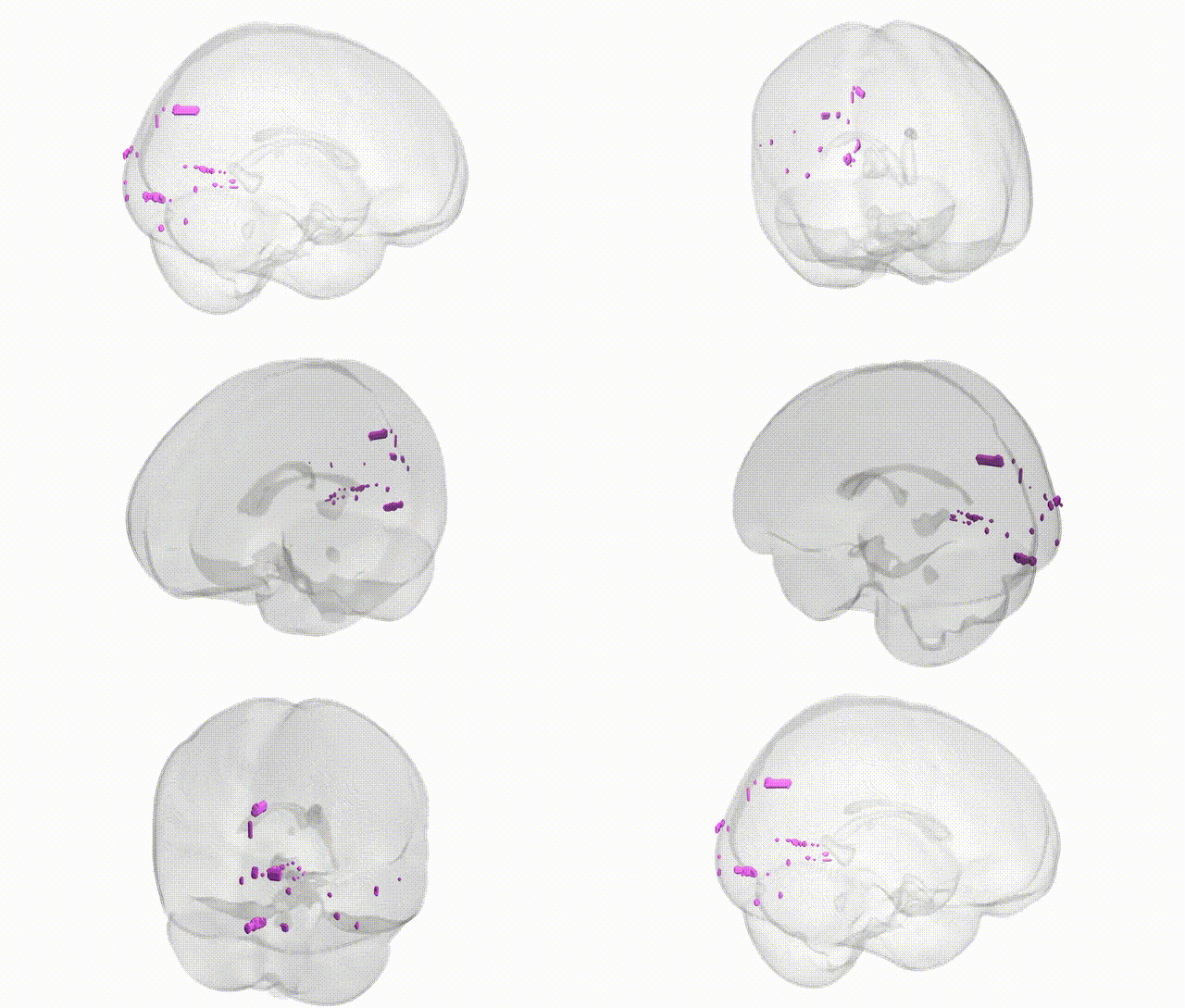
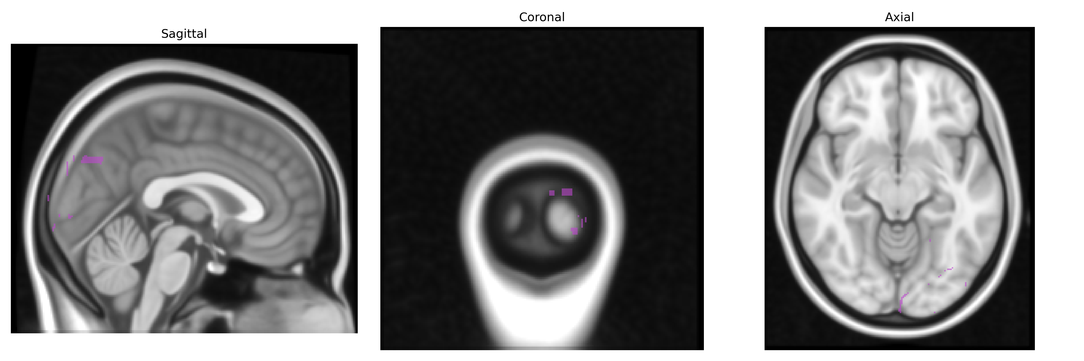
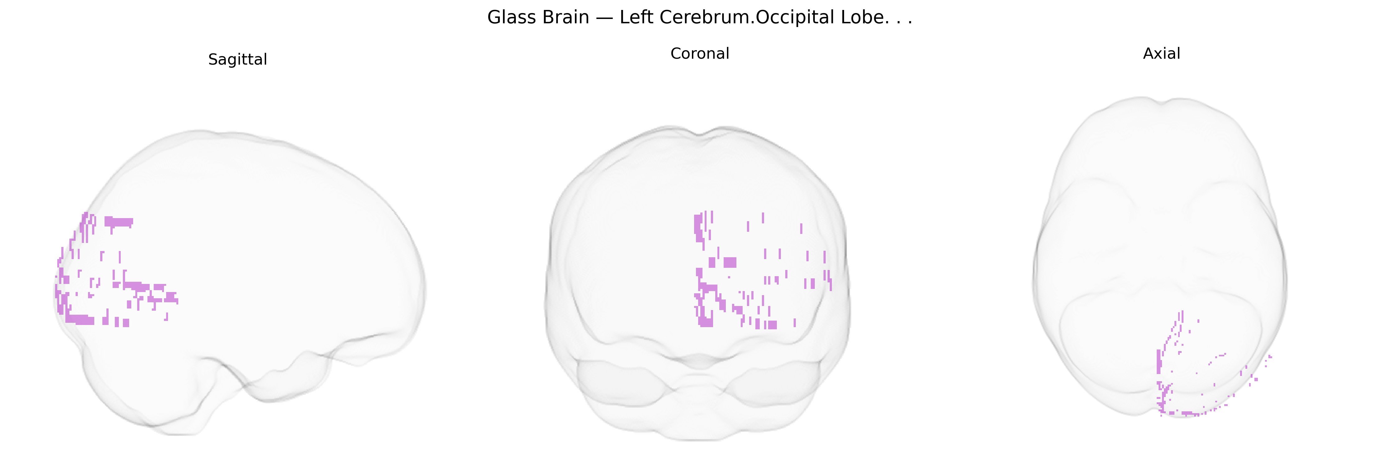
The bilateral Left Cerebrum.Occipital Lobe in the Talairach 1 mm atlas refers to cortical tissue located in the posterior portion of the left cerebral hemisphere, encompassing primary and secondary visual areas that process incoming visual information from the right visual field. This lobe includes regions such as the calcarine cortex (primary visual cortex, or V1) and adjacent extrastriate areas involved in analysis of basic visual features (e.g., orientation, contrast, motion) and higher-order aspects of vision such as form and spatial relationships. Functionally, the left occipital lobe contributes to visual perception, visual recognition, and integration of visual input with language-related processes in nearby temporal and parietal regions.
The bilateral left cerebrum occipital lobe, encompassing primary and associative visual areas in Talairach space, has been implicated in multiple genetic studies linking variation in neurodevelopmental, synaptic, and myelination-related genes to occipital morphology, function, and disease vulnerability. Genome-wide association studies of brain imaging phenotypes (e.g., UK Biobank MRI cohorts) have identified loci in genes such as MAPT, TMEM106B, and multiple microtubule, axon guidance, and cell-adhesion pathways associated with occipital cortical thickness, surface area, and volume, as well as broader “visual network” connectivity. Occipital lobe structural and functional alterations, including in the left hemisphere, have been reported in genetic epilepsies (e.g., occipital lobe epilepsies with implicated genes in ion channels and synaptic transmission), migraine with aura (where polygenic risk overlaps with vascular and neuronal excitability genes), and neurodevelopmental/psychiatric conditions such as autism spectrum disorder and schizophrenia, in which common and rare variants affecting synaptic, GABAergic, and glutamatergic signaling have been associated with altered occipital activation and visual processing. Heritable occipital changes are also described in monogenic and polygenic forms of neurodegeneration (e.g., posterior cortical atrophy in Alzheimer disease risk gene carriers, and C9orf72- or GRN-related frontotemporal lobar degeneration), as well as in disorders with visual field loss or cortical blindness where Mendelian mutations affect visual pathway development. Overall, genetic associations involving the occipital lobe typically act through widespread pathways influencing cortical development, synaptic function, and network connectivity rather than occipital-specific genes, and the Talairach-defined bilateral left occipital region is frequently captured as part of larger, genetically influenced visual and dorsal stream networks in imaging–genetics and GWAS-of-brain-structure studies.
Overview generated by GPT-4o (2026).
Region ID: 246
Hemisphere: bilateral
Atlas: Talairach labels 1mm
Full Quality Version: Download MP4
Full Quality Version: Download MP4
Wali Sidiqyar*, Gaurav Rudravaram*, Elyssa M. McMaster, Trent M. Schwartz, Adam M. Saunders, Kurt G. Schilling, Bennett A. Landman "Introducing SPINS: A Shared Public Visualization Library of Neuroanatomical Structures." Medical Imaging with Deep Learning- short paper
This resource is licensed under CC0 1.0 Universal (Public Domain).